


Tönnis classification
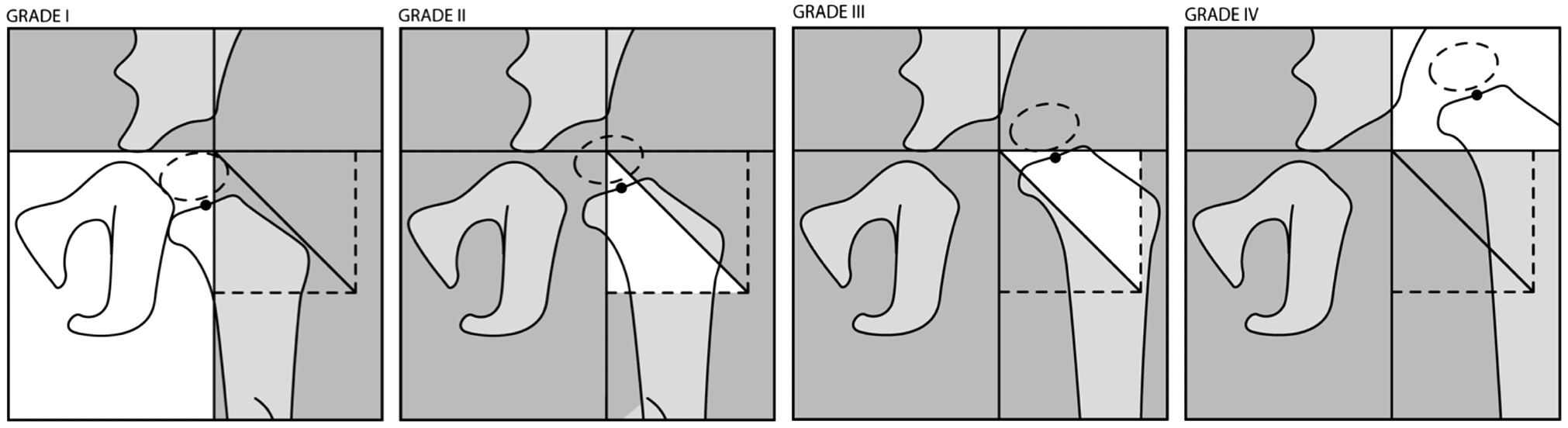
Tönnis classification is a four-grade radiographic system used to describe the severity of femoral-head displacement in developmental dysplasia of the hip (DDH). Grading is based on the position of the femoral-head ossific nucleus relative to Perkins line and the superolateral acetabular margin on an AP pelvic radiograph.
The Tönnis classification provides a simple description of how far the femoral head has displaced from the acetabulum in DDH. It is applied to an appropriately positioned AP pelvis once the femoral-head ossific nucleus is sufficiently visible.
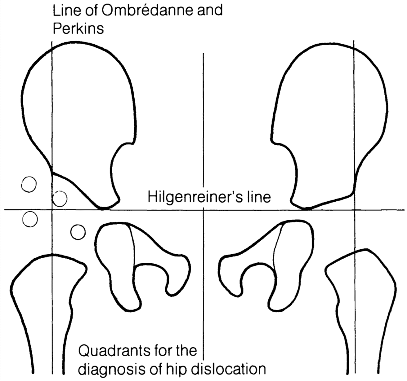
Two principal landmarks are used:
- Perkins line: a vertical line through the lateral margin of the acetabulum.
- Superolateral acetabular margin: the horizontal level of the lateral acetabular roof; later literature often represents this as the superior margin of acetabulum (SMA) line.
| Grade | Position of femoral-head ossific nucleus |
|---|---|
| I | Medial to Perkins line |
| II | Lateral to Perkins line but below the superolateral acetabular margin |
| III | At approximately the level of the superolateral acetabular margin |
| IV | Clearly above the superolateral acetabular margin |
The classification therefore describes increasing superior and lateral displacement, rather than acetabular dysplasia itself. It can be used to standardise the radiographic severity of dislocation and has also been studied as a marker of treatment difficulty and prognosis.
A major limitation is its dependence on a visible femoral-head ossific nucleus. Ossification may be absent, delayed or eccentric in younger children with DDH, making the centre difficult or impossible to define reliably. Severe acetabular dysplasia may also make the lateral acetabular margin difficult to identify.
International Hip Dysplasia Institute grading system 2015
In 2015, Narayanan and colleagues introduced the International Hip Dysplasia Institute (IHDI) classification specifically to overcome the dependence of Tönnis grading on the femoral-head ossific nucleus. Instead, it uses the proximal femoral metaphysis as the reference landmark.
- H-line: Hilgenreiner line through the triradiate cartilages.
- P-line: Perkins line perpendicular to H-line at the superolateral acetabular margin.
- D-line: a new 45° diagonal from the H/P intersection.
- H-point: midpoint of the superior margin of the ossified proximal femoral metaphysis.
History
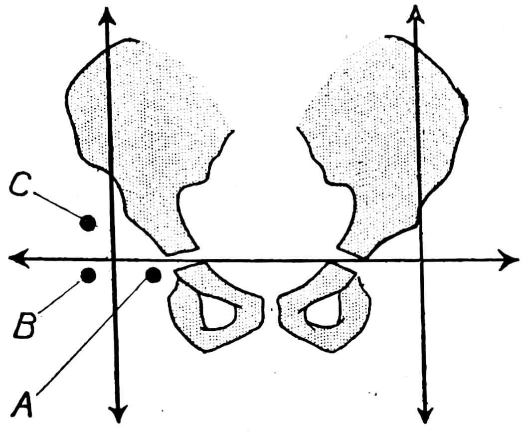
1923 – Louis Ombrédanne (1871-1956) described a horizontal–vertical radiographic construction for localising the femoral-head ossific nucleus in congenital hip dislocation. His illustration showed progressive displacement from the normal inferomedial position (A) to lateral (B) and superolateral (C) positions.
1928 – George Perkins described the vertical acetabular reference through the lateral acetabular margin. His line was incorporated into both quadrant localisation and later displacement grading.
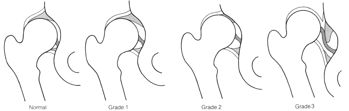
1960s – Becket Howarth (1960) and Peter M Dunn (1969) developed anatomical and pathological grading concepts for congenital hip displacement. Dunn’s necropsy studies demonstrated a progressive spectrum from unstable, dislocatable hips to established dislocation
- Grade I: essentially located but readily dislocatable
- Grade II: increasingly abnormal, commonly partially or completely displaced at rest
- Grade III: head displaced upward/backward with major limbus/capsular changes.
1978 – As arthrograms were unavailable for retrospective evaluation, the Commission for the Study of Hip Dysplasia (Arbeitskreis für Hüftdysplasie) developed a grading system based on the relationship of the ossific centre to acetabular landmarks. Dietrich Tönnis and CSHD adopted a four-grade radiographic system based on the position of the femoral-head ossific nucleus relative to the Ombrédanne–Perkins line and the superolateral acetabular rim. This system was used to standardise severity grading across a large cohorty of treatment-related femoral-head necrosis.
- Grade I – ossific nucleus medial to the Ombrédanne–Perkins vertical
- Grade II – lateral to the vertical but below the superolateral acetabular rim
- Grade III – midpoint approximately level with the superolateral rim
- Grade IV – clearly above the superolateral rim
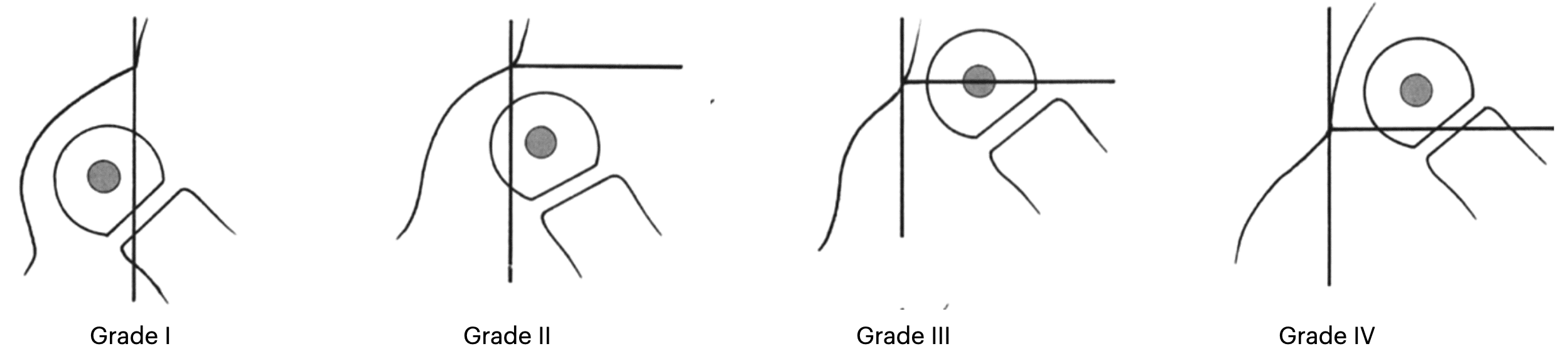
Tonnis et al. 1978
1982 – The classification was reproduced in the English-language collective report Congenital hip dislocation – avascular necrosis, prepared by the Commission for the Study of Hip Dysplasia.
1984 – Dietrich Tönnis reproduced the CSHD grading system in his major German and English monographs and distinguished it from the older Hilgenreiner + Ombrédanne-Perkins quadrant method used for general localisation. Through these texts the working-group system became increasingly associated with Tönnis’s name.
2015 – Narayanan et al. introduced the International Hip Dysplasia Institute (IHDI) classification to overcome the dependence of Tönnis grading on a visible femoral-head ossific nucleus. The ossific centre was replaced by the H-point at the proximal femoral metaphysis. Hilgenreiner and Perkins lines were retained and a new 45° diagonal line was introduced.
Eponymythology
The Tönnis classification was originally a collaborative grading system rather than the sole creation of Dietrich Tönnis. In 1984, Tönnis attributed the four-grade radiographic system to the Arbeitskreis für Hüftdysplasie / Commission for the Study of Hip Dysplasia of the German Society for Orthopaedics and Traumatology. He cited publications from 1978 and 1982. Through Tönnis’s subsequent textbooks and publications, the system became widely known as the Tönnis classification.
The radiographic foundations were older still. Hilgenreiner, Ombrédanne and Perkins had already established reference lines and quadrants for localising the femoral head. The working group’s contribution was to convert displacement relative to these landmarks into a standardised four-grade radiographic severity system
Associated Persons
- Heinrich Hilgenreiner (1870–1953)
- Louis Ombrédanne (1871-1956)
- George Perkins (1892–1979)
- Dietrich Tönnis (1927-2010)
Alternative names
- Tönnis method
References
Historical references
- Howarth MB. Congenital dislocation of the hip is not a primary dysplasia of the acetabulum. 8th International Kongress SICOT, Rapport. Imprimerie des Sciences, Bruxelles, 1960: 150-162
- Dunn PM. Congenital dislocation of the hip (CDH): necropsy studies at birth. Proc R Soc Med. 1969 Oct;62(10):1035-7.
- Dunn PM. Perinatal observations on the etiology of congenital dislocation of the hip. Clin Orthop Relat Res. 1976 Sep;(119):11-22.
- Tönnis D. Hüftluxation und Hüftkopfnekrose. Eine Sammelstatistik des Arbeitskreises für Hüftdysplasie der DGOT (Deutsche Gesellschaft für Orthopädische Traumatologie), Bücherei des Orthopäden, Bd.21. Enke, Stuttgart 1978
- Tönnis D, et al. Congenital hip dislocation – avascular necrosis. Collective statistics prepared by the Commission for the Study of Hip Dysplasia. Thieme-Stratton, New York, 1982.
- Tönnis D. Die angeborene Hüftdysplasie und Hüftluxation im Kindes- und Erwachsenenalter. 1984
- Tönnis D. Congenital Dysplasia and Dislocation of the Hip in Children and Adults. 1987
Eponymous term review
- Narayanan U, Mulpuri K, Sankar WN, Clarke NM, Hosalkar H, Price CT; International Hip Dysplasia Institute. Reliability of a New Radiographic Classification for Developmental Dysplasia of the Hip. J Pediatr Orthop. 2015 Jul-Aug;35(5):478-84
- Terjesen T, Horn J. Prognostic value of severity of dislocation in late-detected developmental dysplasia of the hip. J Child Orthop. 2020 Aug 1;14(4):266-272.
- Shapira J, Chen JW, Bheem R, Rosinsky PJ, Maldonado DR, Lall AC, Domb BG. Binary Tönnis classification: simplified modification demonstrates better inter- and intra-observer reliability as well as agreement in surgical management of hip pathology. BMC Musculoskelet Disord. 2020 Jul 29;21(1):502
- Karnik A, Lawande A, Lawande MA, Patkar D, Aroojis A, Bhatnagar N. Practice Essentials of Imaging in Early Diagnosis of DDH. Indian J Orthop. 2021 Oct 7;55(6):1466-1479.
- Kyle E, Cadogan M. Hip and pelvic lines and arcs. LITFL
eponymictionary
the names behind the name

BA MA (Oxon) MBChB (Edin) FACEM FFSEM. Emergency physician, Sir Charles Gairdner Hospital. Passion for rugby, medical history, medical education, and asynchronous learning #FOAMed evangelist. Editor-in-Chief and Webmaster of Life in the Fast lane, LITFL | On Call 4e| Eponyms | Books | Horology |