![]()
De Winter T Wave
First reported by de Winter in 2008, the de Winter ECG pattern is an anterior STEMI equivalent that presents without obvious ST segment elevation

![]()
First reported by de Winter in 2008, the de Winter ECG pattern is an anterior STEMI equivalent that presents without obvious ST segment elevation

Heart HQ - Episode 3: Valve-in-Valve Transcatheter Aortic Valve Implantation (ViV TAVI)and Cardiovascular Disease in women
A single agent overdose causing AV blockade, QRS widening, and QT prolongation.... but reports of death only if QRS > 200ms. Which medication is this?

Internet Archive founder, Brewster Kahle, reflects back on the most surprising advancement of his early innovation, the Wayback Machine.

Sir Peter James Kerley (1900-1979) was an Irish radiologist. Kerley was widely published including describing (but not naming) his eponymous lines firstly in 1933 and then in again his textbook in 1950, and widely about TB diagnosis. Kerley lines A, B and C
Debrief: Following SMACCForce Simulation Team of a Resuscitative Hysterotomy in a high-pressured situation.
SIM team demonstrate a Resuscitative Hysterotomy/ Perimortem Caesarean section on a pregnant female involved in a high speed motor vehicle accident.
Serotonin syndrome results from drug-induced over-stimulation of serotonin receptors in the CNS and is characterized by a triad of CNS dysfunction, autonomic disturbance and neuromuscular effects; aka serotonin toxicity
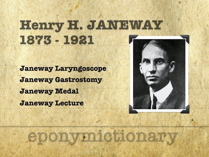
Henry Harrington Janeway (1873-1921) was an American physician and pioneer for radiation therapy in the treatment of cancer. Janeway Gastrostomy, Janeway Laryngoscope
Heart HQ - Episode 2: Wolff-Parkinson-White (WPW) Syndrome, myocarditis and COVID 19 Vaccines
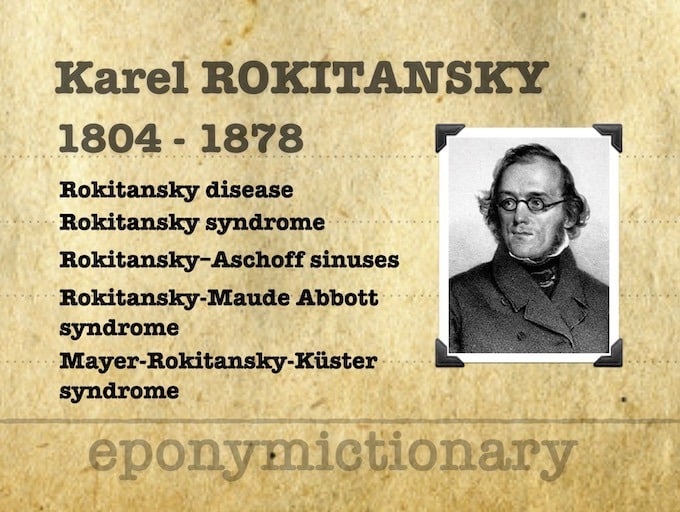
Karel (Carl von) Rokitansky (1804-1878) was a Czech pathologist. Eponymous terms include Rokitansky disease; Rokitansky syndrome; Rokitansky–Aschoff sinuses; and Rokitansky-Maude Abbott syndrome

Pick a body part. Pick a skin surface. Pick a block. The Nerve Block App makes regional anaesthesia easier on the go