![]()
Adie syndrome
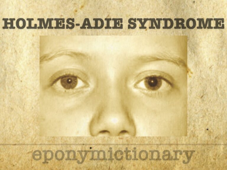
Holmes–Adie syndrome: a benign neurological condition marked by tonic pupils and areflexia, historically mistaken for neurosyphilis.

![]()
Holmes–Adie syndrome: a benign neurological condition marked by tonic pupils and areflexia, historically mistaken for neurosyphilis.

Cornelia Catharina de Lange (1871-1950) was a Dutch pediatrician. Described Cornelia de Lange syndrome (CdLS) in 1933
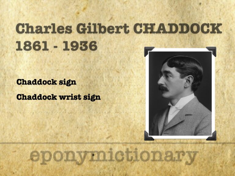
Charles Gilbert Chaddock (1861-1936) was an American neurologist, psychiatrist, poet and medical translator. Chaddock sign and Chaddock wrist sign
Emergency Procedure, instruction and discussion: nail bed repair - a deceptively simple injury that can lead to permanent nail deformity if managed incorrectly

Józef Dietl (1804–1878) was a Polish physician, politician, professor and rector. Eponym: Dietl's crisis ureteropelvic junction obstruction (UPJO)
Emergency Procedure: nail bed lacerations - a deceptively simple injury that can lead to permanent nail deformity if not managed carefully
Leslie Zieve (1915–2000), American hepatologist; Zieve syndrome: jaundice, haemolysis, and hyperlipidaemia in fatty liver disease.
Leonardo Gigli (1863-1908) was an Italian surgeon and gynaecologist. Inventor of the Gigli saw for lateralised pubiotomy
Emergency Procedure, instruction and discussion: Serratus Anterior Plane (SAP) Block; a technique most often used for rib fracture pain
Emergency Procedure: Serratus Anterior Plane (SAP) Block; a technique most often used for rib fracture pain
Neurocysticercosis. Third edition in our Neuroimaging case study series with guest editors Drs. Michael Leonard and David Weinrib

Edmund Landolt (1846–1926): Swiss-French ophthalmologist who created the Landolt C optotype, advanced strabismus surgery, and retinal anatomy studies