![]()
Echo basics: Measurements and Reports
Echocardiography and valve measurements. Comprehensive assessment requires measurements to be made from 2D images and the waveforms generated during Doppler investigations

![]()
Echocardiography and valve measurements. Comprehensive assessment requires measurements to be made from 2D images and the waveforms generated during Doppler investigations
Neuro 101: Cerebral Hemispheres. Clinicoanatomic correlation for frontal, temporal, parietal and occipital lobes. Overview of anterior and posterior arterial circulation

Non-traumatic abdominal ecchymosis of the abdominal wall and flanks (Grey Turner, Cullen and Stabler); scrotum (Bryant) and upper thigh (Fox) as clues to potentially serious causes of abdominal pathology.
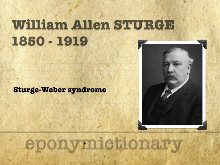
William Allen Sturge (1850–1919) English neurologist and archaeologist; first described Sturge-Weber syndrome; awarded MVO; pioneer of women’s medical education; noted collector of prehistoric artefacts.

Corneal foreign bodies present with pain, watering, and irritation. Remove under anaesthesia, exclude penetrating injury, and arrange follow-up.
Bacterial conjunctivitis is common and treatable, but screen for serious infections like gonococcus, meningococcus, and trachoma in high-risk patients.
Acute loss of vision is an ophthalmic emergency. Assess urgently. Persistent or unexplained cases require immediate specialist ophthalmology input.
Acute non-traumatic loss of vision is an ophthalmic emergency. All patients require urgent assessment, and persistent deficits mandate immediate ophthalmology referral.
Chemical eye injuries are emergencies. Immediate irrigation, category 2 triage, and ophthalmology input are critical to preserve vision and minimise damage.
Neuro 101: Neurological Examination. The eight steps, mental status, motor, sensory, reflex, cerebellar examinations
Echocardiography and valve views. Overview of valve disease and parasternal, apical and subcostal valve views with the echo probe
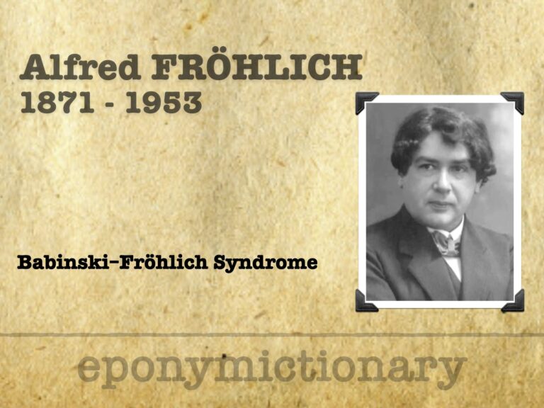
Alfred Fröhlich (1871-1953) Austrian neurologist and pharmacologist; pioneer of neuroendocrinology who described adiposogenital dystrophy, linking pituitary lesions to obesity and hypogonadism.