![]()

John Henry Bryant
John Henry Bryant (1867–1906) English physician. Eponym: Blue Scrotum Sign of Bryant associated with ruptured abdominal aortic anurysm (1903)

![]()
John Henry Bryant (1867–1906) English physician. Eponym: Blue Scrotum Sign of Bryant associated with ruptured abdominal aortic anurysm (1903)

Non-traumatic abdominal ecchymosis of the abdominal wall and flanks (Grey Turner, Cullen and Stabler); scrotum (Bryant) and upper thigh (Fox) as clues to potentially serious causes of abdominal pathology.

Overview of Abdominal Aortic Aneurysm: presentation, risk factors, rupture risk, clinical features, investigations, emergency management, and surgical options
Abdominal Aortic Aneurysm (AAA) Surveillance Chart. All incidentally found aortic aneurysms should be referred to a vascular surgeon if the patient is a potential candidate for surgery.

Bryant’s sign: Scrotal ecchymosis associated with ruptured abdominal aortic aneurysm (AAA) first described in 1903 by John Henry Bryant (1867-1906)
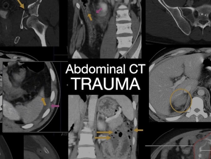
Abdominal CT: Trauma series. Solid organ injuries. Trauma is a leading cause of death worldwide and it has two broad classifications: Blunt and penetrating
Abdominal CT: Identifying intestinal ischaemia. Mesenteric ischaemia can be visualised on CT through examining blood vessels and the bowel

POCUS made easy: AAA scan is useful to look for a ruptured or leaking abdominal aortic aneurysm

An 80-year-old man presents with acute abdominal pain. He is in a shocked state, HR 95, BP 82/55, peripherally shutdown.

Grey Turner sign refers to bruising of the flanks. Originally described 1919 (published 1920) by George Grey Turner (1877–1951) most commonly associated with acute pancreatitis

Fox's sign: non-traumatic ecchymosis over the upper outer aspect of the thigh secondary to abdominal haemorrhage. First described by English surgeon John Adrian Fox in 1966
John Adrian Fox English surgeon. Eponym: Fox's sign (1966) non-traumatic ecchymosis upper outer thigh with abdominal haemorrhage