![]()
Abdominal CT: cholecystitis
Imaging cholecystitis
Cholecystitis is inflammation of the gallbladder commonly caused by an obstruction of the cystic duct by a gallstone. Clinically it typically presents with right upper quadrant pain, elevated biliary enzymes and an elevated white blood cell count
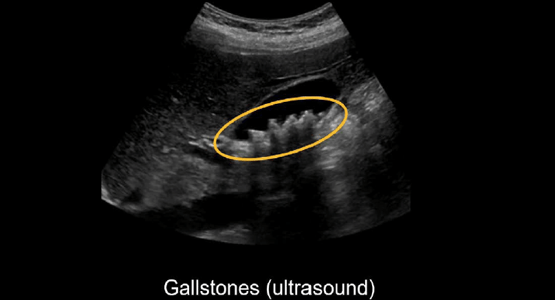
Ultrasound is often the first study done for right upper quadrant pain and it is great at looking for gallstones.
Even though ultrasound is the preferred imaging study for diagnosing acute cholecystitis, it is not uncommon to encounter this condition on CT. This is because acute cholecystitis can mimic other causes of abdominal pain.
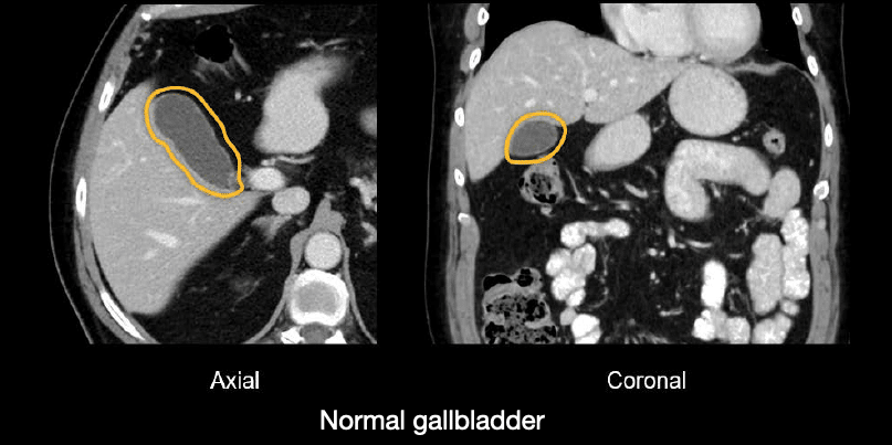
The good news is that CT is quite good at making this diagnosis. On CT, the normal gallbladder has a thin wall with clean, dark fat surrounding it, and it looks like it is filled with fluid.
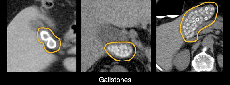
It is fairly common to encounter gallstones on CT, particularly when they are calcified and have a bright appearance. Here are some examples of gallstones of various sizes and number.
Key findings
When diagnosing acute cholecystitis, there are three key CT imaging findings
- Dilated gallbladder
- Thick, irregular gallbladder wall
- Stranding and fluid
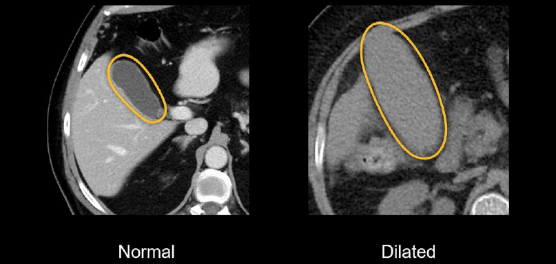
Dilated gallbladder When the gallbladder is obstructed, it dilates, and it will look like an inflated balloon compared to the normal gallbladder.
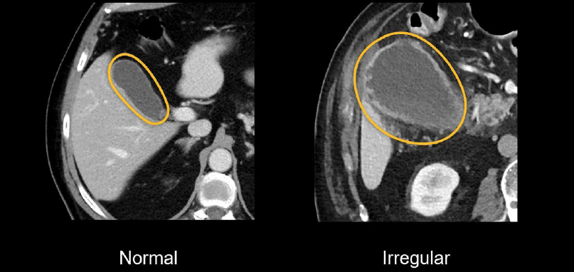
Thick, irregular gallbladder wall The gallbladder wall will become thick and is often irregular when inflammation is present. The normal gallbladder wall is a thin, bright line in comparison.
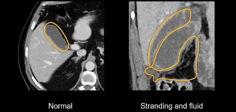
Stranding and fluid Acute cholecystitis will result in fluid and stranding of the surrounding fat due to inflammation.
Knowledge iteration
Scroll through these PACS viewer images
- Scroll down to the gallbladder.
- The first thing you’ll notice is that the gallbladder is elongated and dilated. There is also subtle stranding near the gallbladder neck, and the fundus is displacing the adjacent hepatic flexure inferiorly.
- Notice how the gallbladder pushes against the anterior abdominal wall, slightly indenting it. What does that tell us about the gallbladder?
- The gallbladder must be quite firm to indent the abdominal wall rather than conforming to it, and that can only happen if the gallbladder is obstructed and under pressure. This is called the tensile fundus sign.
- The tensile fundus sign, along with the surrounding stranding and inflammation, can help us confidently make the diagnosis of acute cholecystitis.
This is an edited excerpt from the Medmastery course Abdominal CT Pathologies by Michael P. Hartung, MD. Acknowledgement and attribution to Medmastery for providing course transcripts
- Hartung MP. Abdominal CT: Common Pathologies. Medmastery
- Hartung MP. Abdominal CT: Essentials. Medmastery
- Hartung MP. Abdomen CT: Trauma. Medmastery
References
- Top 100 CT scan quiz. LITFL
- Rippey J. Ultrasound Case 049. LITFL
Radiology Library: Acute abdomen. Solid organ and Vascular pathology
- Hartung MP. Abdominal CT: acute interstitial pancreatitis
- Hartung MP. Abdominal CT: acute necrotizing pancreatitis
- Hartung MP. Abdominal CT: renal stones and the flank pain CT
- Hartung MP. Abdominal CT: renal infections
- Hartung MP. Abdominal CT: cholecystitis
- Hartung MP. Abdominal CT: intestinal ischaemia
- Hartung MP. Abdominal CT: rupturing abdominal aortic aneurysm
Abdominal CT interpretation

Assistant Professor of Abdominal Imaging and Intervention at the University of Wisconsin Madison School of Medicine and Public Health. Interests include resident and medical student education, incorporating the latest technology for teaching radiology. I am also active as a volunteer teleradiologist for hospitals in Peru and Kenya. | Medmastery | Radiopaedia | Website | Twitter | LinkedIn | Scopus

MBChB (hons), BMedSci - University of Edinburgh. Living the good life in emergency medicine down under. Interested in medical imaging and physiology. Love hiking, cycling and the great outdoors.