![]()
Abdominal CT: genitourinary system
Imaging the genitourinary system
Kidneys
The kidney has a smooth, bean-like shape, normally measuring 9–14 cm in length. The normal kidney will enhance uniformly. However, depending on the phase of contrast, you might be able to differentiate between the outer renal cortex and inner medulla, as shown below.
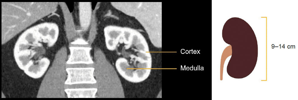
On a portal venous phase CT, the outer renal cortex is usually brighter than the inner, triangular-shaped medulla which points toward the renal pelvis. It is generally easiest to evaluate the kidney on coronal images where you can review them in their long axis, and it is easier to compare them side-by-side.
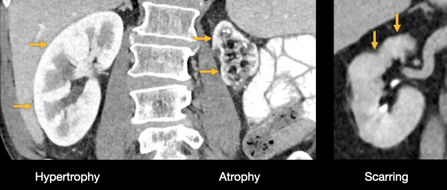
Scarring of the kidney can happen after infection, blockage of the blood supply, or traumatic injury. It causes the kidney to lose volume and appear irregular in shape.
Atrophic (i.e., abnormally small kidneys) are commonly seen in the setting of severe trauma or chronic renal failure. The next image shows an example of an atrophic left kidney several years after a major vascular injury. Notice how the right kidney appears larger than normal, becoming hypertrophic. This occurred over time to compensate for the loss of function of the injured left kidney
Masses
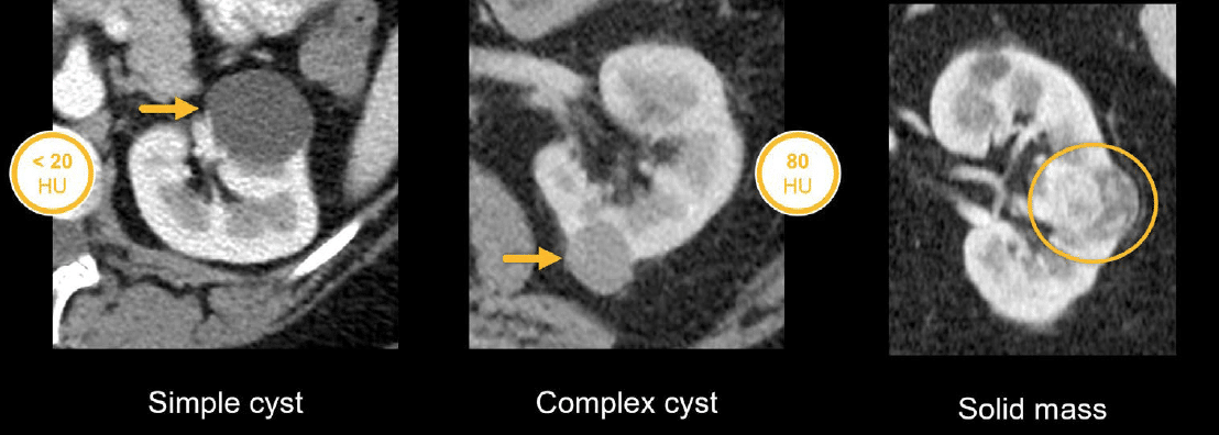
There are a number of different types of masses that you may encounter when evaluating the kidneys. The most common renal masses are benign simple cysts which become increasingly common as patients get older.
The simple cyst will typically look dark, much like fluid, and will measure less than 20 Hounsfield units (HU). However, a complex cyst containing protein or blood products will appear bright or dense. For example, the complex cyst shown below measures 80 HU.
Solid, enhancing masses may appear irregular as in the example below. These are concerning for renal cell carcinoma and require further evaluation and management by urology.
Ureters
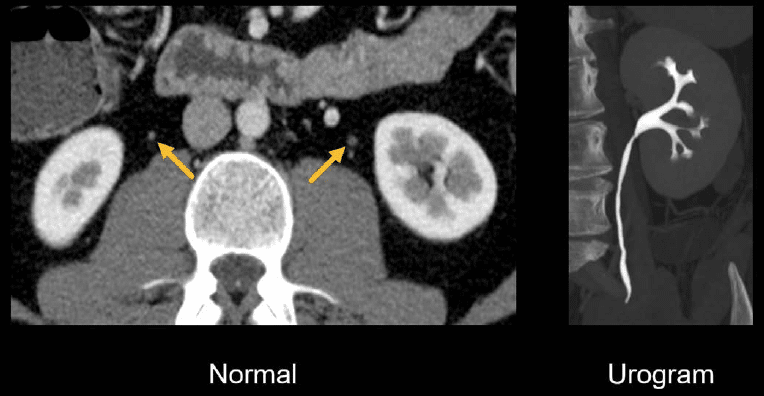
From the kidneys, you can follow the ureters down to where they drain into the bladder.
Normal ureters have a thin wall and small diameter, which is why they are best evaluated on a CT urogram where they are filled with contrast-mixed urine and easier to see. This is especially true in very thin patients—it can be difficult to follow normal ureters in these patients as the ureters are often pressed against surrounding structures.
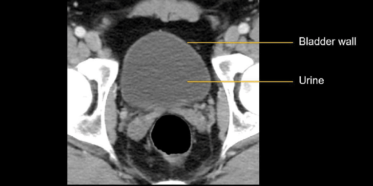
Bladder
Like the ureter, the wall of a normal urinary bladder is relatively thin when distended with urine, which looks like simple fluid.
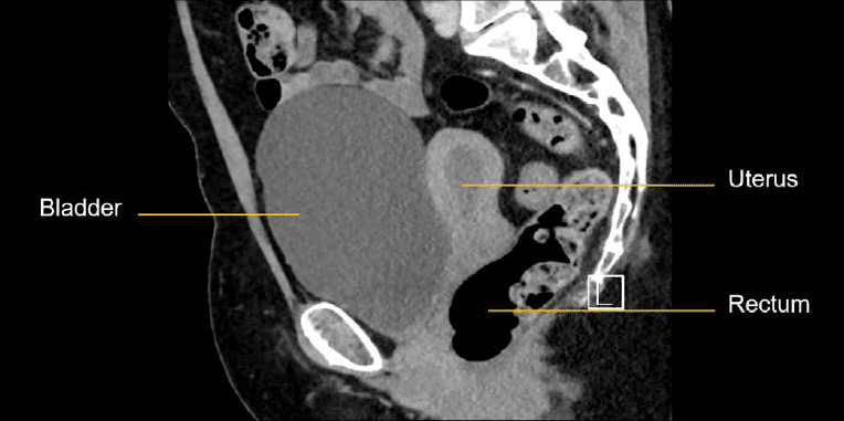
It can be helpful to evaluate the bladder on sagittal images where you can see it relative to the other major pelvic structures. In the CT images from a female patient, shown below, we can evaluate the bladder, uterus, and rectum in their long axes.
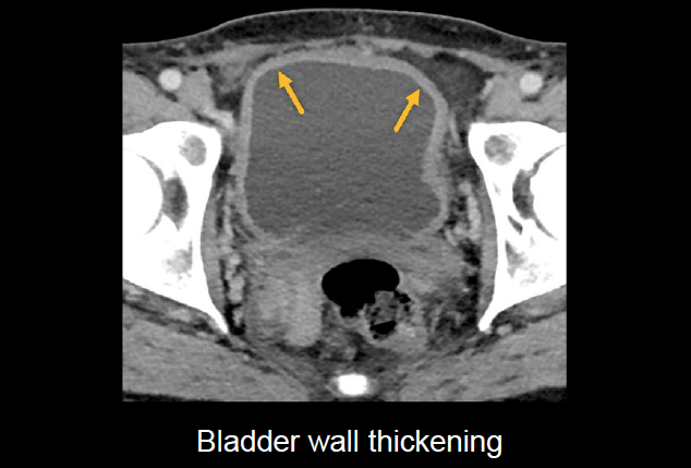
The bladder wall can become thickened due to infection, inflammation, a tumour, or chronic outflow compromise. Often the patient’s history, symptoms, and urinalysis are needed to determine the cause of wall thickening.
Reproductive organs
CT is not the best method to evaluate the reproductive organs; this is better accomplished with ultrasound or magnetic resonance imaging (MRI). However, we can still sometimes catch important or obvious abnormalities with CT.
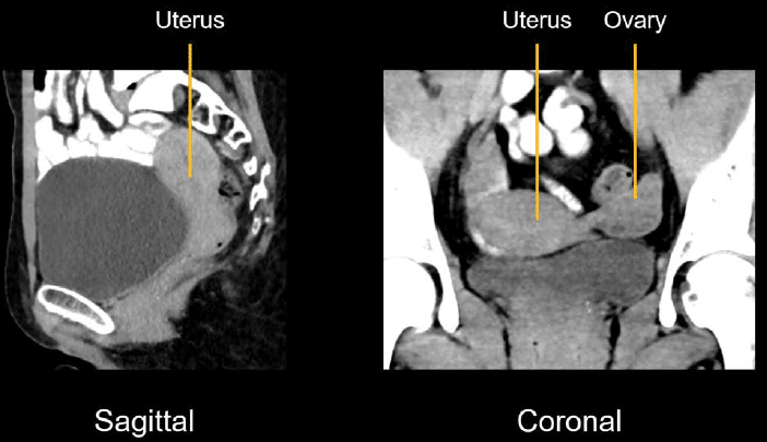
Uterus and ovaries
The female reproductive organs seen on CT include the uterus and ovaries. The vagina is usually collapsed and not well seen on CT. The uterus is usually easiest to see on the sagittal view, whereas the ovaries are best seen on either the axial or coronal view.
Note: the ovaries are easier to identify in younger patients since they atrophy after menopause, becoming smaller.
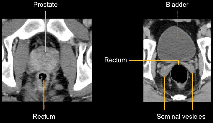
Prostate and seminal vesicles
In males, the prostate and seminal vesicles can be evaluated for normal size and shape, fluid collections and surrounding inflammation, or obvious masses. The axial images featured next show their relationship with the bladder and rectum.
Knowledge iteration
To solidify the key anatomy from this lesson, click on the image below to scroll through a set of images which show healthy-appearing kidneys, bladder, prostate, and seminal vesicles on a male patient.
- Starting with the axial images, notice that the kidneys enhance normally and symmetrically with increased enhancement of the outer cortex in comparison to the inner medulla.
- The vascular supply is open, and you can see the renal vein and arteries.
- You can follow the collecting system down to the ureter and follow the ureter to the urinary bladder.
- At the urinary bladder, there is a normal, thin wall, and there are normal contours of the prostate and seminal vesicles.
- The coronal images provide a view of the long axis of the kidneys making for easy side-by-side comparison.
- In the pelvis, you can appreciate the relationship between the urinary bladder as it sits on the prostate.
This is an edited excerpt from the Medmastery course Abdominal CT Essentials by Michael P. Hartung, MD. Acknowledgement and attribution to Medmastery for providing course transcripts.
- Hartung MP. Abdominal CT: Common Pathologies. Medmastery
- Hartung MP. Abdominal CT: Essentials. Medmastery
- Hartung MP. Abdomen CT: Trauma. Medmastery
References
- Top 100 CT scan quiz. LITFL
Radiology Library: Abdominal CT: Imaging the abdominal organs and bowel
- Hartung MP. Abdominal CT: Evaluating the liver
- Hartung MP. Abdominal CT: Evaluating the biliary system and pancreas
- Hartung MP. Abdominal CT: Imaging the spleen and adrenal glands
- Hartung MP. Abdominal CT: Mastering the genitourinary system
- Hartung MP. Abdominal CT: Evaluating the lower oesophagus and stomach
- Hartung MP. Abdominal CT: Imaging the small bowel
- Hartung MP. Abdominal CT: Viewing the peritoneal cavity
- Hartung MP. Abdominal CT: Running the large bowel and appendix
Abdominal CT interpretation

Assistant Professor of Abdominal Imaging and Intervention at the University of Wisconsin Madison School of Medicine and Public Health. Interests include resident and medical student education, incorporating the latest technology for teaching radiology. I am also active as a volunteer teleradiologist for hospitals in Peru and Kenya. | Medmastery | Radiopaedia | Website | Twitter | LinkedIn | Scopus

MBChB (hons), BMedSci - University of Edinburgh. Living the good life in emergency medicine down under. Interested in medical imaging and physiology. Love hiking, cycling and the great outdoors.