Abdominal CT: spleen and adrenal glands
Imaging the spleen and adrenal glands
Spleen
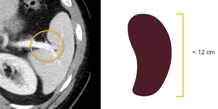
The spleen is less complex than the liver and is usually straightforward to evaluate. It is located in the left upper quadrant and has an oval or wedge shape. It generally measures less than 12 cm in length, but a volume estimate is a more accurate way to determine if the spleen is enlarged.
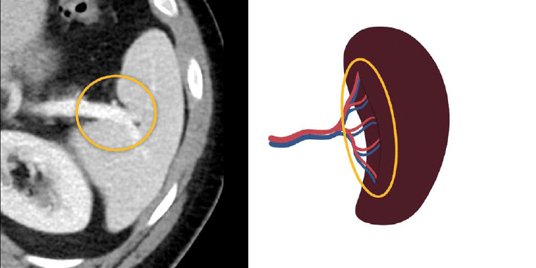
The splenic artery and vein enter the spleen through a central depression called the splenic hilum. The hilum is also associated with ligaments attaching the spleen to the stomach and left kidney, which are not usually visible on CT
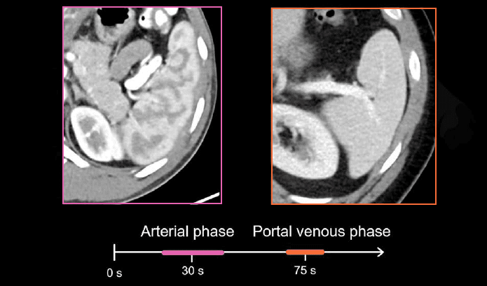
In the arterial phase, the spleen often has a wavy pattern of enhancement that is referred to as a zebra-striped appearance. This undulating pattern of enhancement is caused by the contrast making its way through the splenic tissue. Over time this pattern will become less pronounced, and it eventually disappears by the portal venous phase.
Benign masses
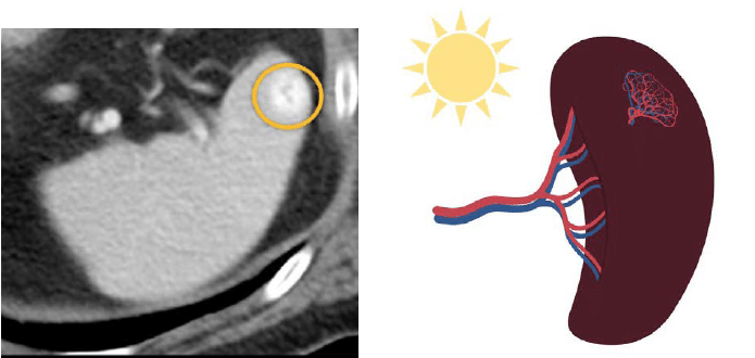
Most lesions in the spleen are benign and incidental, such as cysts or hemangiomas. Cysts do not enhance, and they appear dark compared to the splenic tissue, similar to fluid.
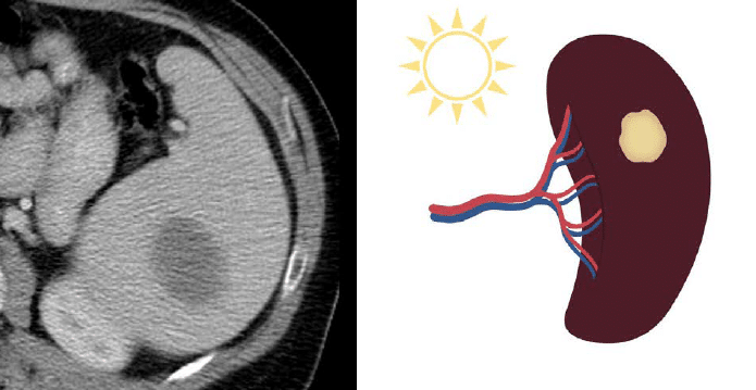
On the other hand, hemangiomas will enhance, and they can be either darker or brighter than the normal splenic tissue.
Malignant masses
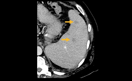
Not all splenic masses are benign. When staging cancer, it is important to carefully evaluate the spleen, as cancers like breast, lung, colorectal, and melanoma can spread there.
Lymphoma commonly involves the spleen and can also cause both an enlarged spleen and masses.
Adrenal glands
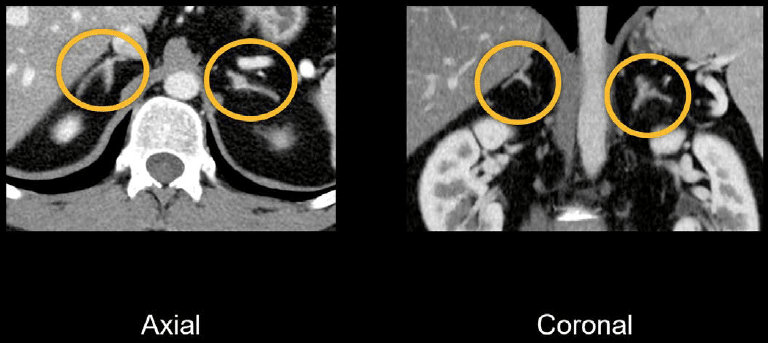
The adrenal glands are paired endocrine organs that are found anterior and superior to the kidneys. The normal adrenal gland is thin and enhances uniformly with a characteristic y-shape.
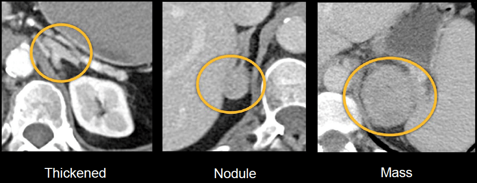
You may see thickened adrenal glands or nodules, which are often benign. However, both primary cancer and metastatic disease can occur in the adrenal glands. There are dedicated adrenal exams for evaluating suspicious nodules.
Knowledge iteration
To solidify the key anatomy from this lesson, click on the image below to scroll through a set of images featuring a healthy-appearing spleen and adrenal glands.
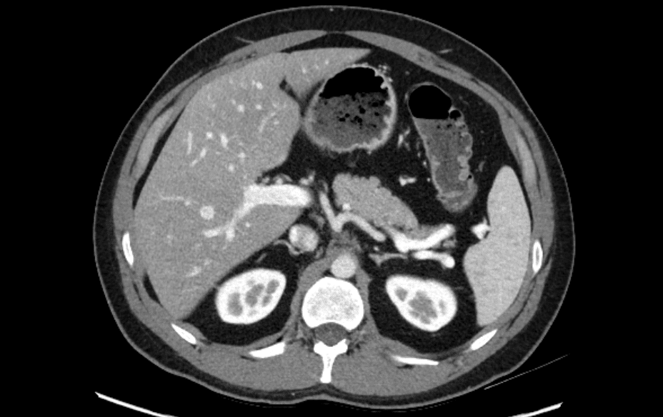
- Looking at the axial images, you can start with the spleen in the left upper quadrant. Notice its smooth contour, normal size, and vascular supply entering the splenic hilum.
- The adrenal glands lie above and slightly in front of both kidneys, with a characteristic y-shape. The adrenal glands are thin, uniformly enhancing, and without a nodule or mass.
- Next, review that same anatomy on the coronal images. The spleen is in the left upper quadrant with the artery and vein entering the splenic hilum.
- The adrenal glands are sitting above the kidneys. See if you can identify the characteristic shape and thin, normal, enhancing parenchyma
This is an edited excerpt from the Medmastery course Abdominal CT Essentials by Michael P. Hartung, MD. Acknowledgement and attribution to Medmastery for providing course transcripts.
- Hartung MP. Abdominal CT: Common Pathologies. Medmastery
- Hartung MP. Abdominal CT: Essentials. Medmastery
- Hartung MP. Abdomen CT: Trauma. Medmastery
References
- Top 100 CT scan quiz. LITFL
Radiology Library: Abdominal CT: Imaging the abdominal organs and bowel
- Hartung MP. Abdominal CT: Evaluating the liver
- Hartung MP. Abdominal CT: Evaluating the biliary system and pancreas
- Hartung MP. Abdominal CT: Imaging the spleen and adrenal glands
- Hartung MP. Abdominal CT: Mastering the genitourinary system
- Hartung MP. Abdominal CT: Evaluating the lower oesophagus and stomach
- Hartung MP. Abdominal CT: Imaging the small bowel
- Hartung MP. Abdominal CT: Viewing the peritoneal cavity
- Hartung MP. Abdominal CT: Running the large bowel and appendix
Abdominal CT interpretation
Assistant Professor of Abdominal Imaging and Intervention at the University of Wisconsin Madison School of Medicine and Public Health. Interests include resident and medical student education, incorporating the latest technology for teaching radiology. I am also active as a volunteer teleradiologist for hospitals in Peru and Kenya. | Medmastery | Radiopaedia | Website | Twitter | LinkedIn | Scopus
MBChB (hons), BMedSci - University of Edinburgh. Living the good life in emergency medicine down under. Interested in medical imaging and physiology. Love hiking, cycling and the great outdoors.


