
![]()
Abdominal CT: peritoneal cavity
Viewing the peritoneal cavity
Duodenum
The peritoneal cavity is the major abdominal cavity containing many of the organs and most of the bowel. We’ll also take a look at the omentum – the fatty, apron-like covering that hangs in front of the small bowel and mesentery.

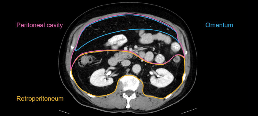
The boundaries of the peritoneum and retroperitoneum are not usually visible on CT unless there is disease or fluid. You often have to infer where they are, based on which organs are normally in each space.
Retroperitoneum
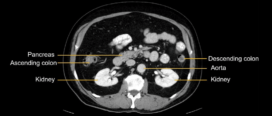
The following structures are located in the retroperitoneum:
- Kidneys
- Aorta
- Pancreas
- Ascending and descending colon
This knowledge helps you to draw a mental boundary around that space. That leaves us with the space in front of the peritoneal cavity, which contains the rest of the bowel, mesentery, and omentum.
Evaluating the peritoneal cavity
When evaluating the peritoneal cavity, you should be looking for the following:
- Nodules or masses in the omentum
- Ascites (i.e., areas where fluid has collected)
- Peritoneal thickening
Nodules or masses in the omentum
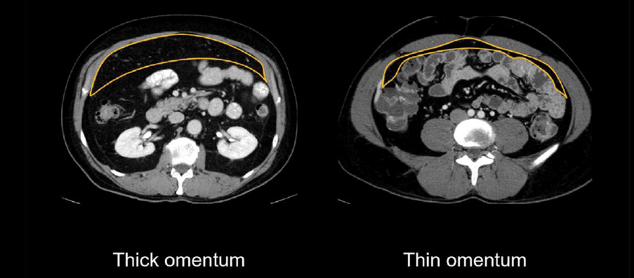
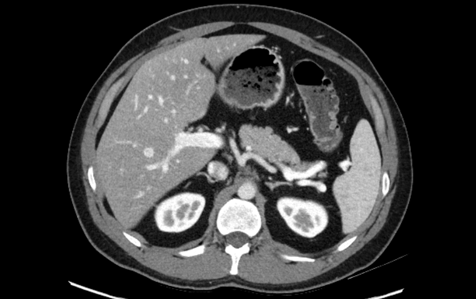
Normally, the omentum blends in with the rest of the peritoneal and mesenteric fat, but you can identify it by looking for fat density with vessels running through it in front of the small bowel.

The omentum can be recognized by the fat density draping over the bowel. Depending on the patient’s intraabdominal fat content, it can be quite thick, as shown in the left-hand image below, or rather thin, as in the right-hand image, barely separating the bowel from the anterior body wall.
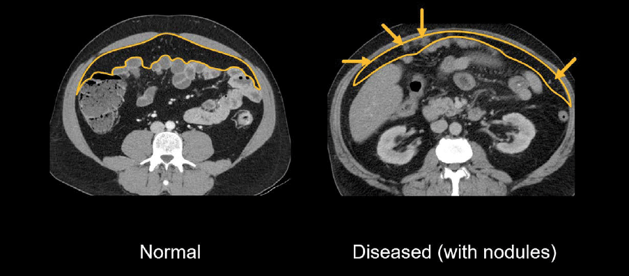
When the omentum is diseased or infiltrated by a tumor, you will often see nodules and thickening, indicating the spread of a tumor within the peritoneal cavity.
In the images below, cancer has spread extensively throughout the peritoneal cavity but with different severities of disease. The example on the left shows relatively subtle nodules in the omentum, whereas in the example on the right, you can see extensive tumor infiltrating the omentum, with surrounding ascites. This is called an omental cake.

Ascites
Ascites can be quantified as small, medium, and large, and is associated with a variety of diseases – most commonly, liver disease.
Ascites can distribute throughout the peritoneal cavity, as it is often a simple fluid that moves and flows freely. When you are checking for fluid on CT, look in the upper, middle, and pelvic regions of the abdomen and pelvis. The distribution of ascites within the abdominal cavity will depend on the quantity and cause.
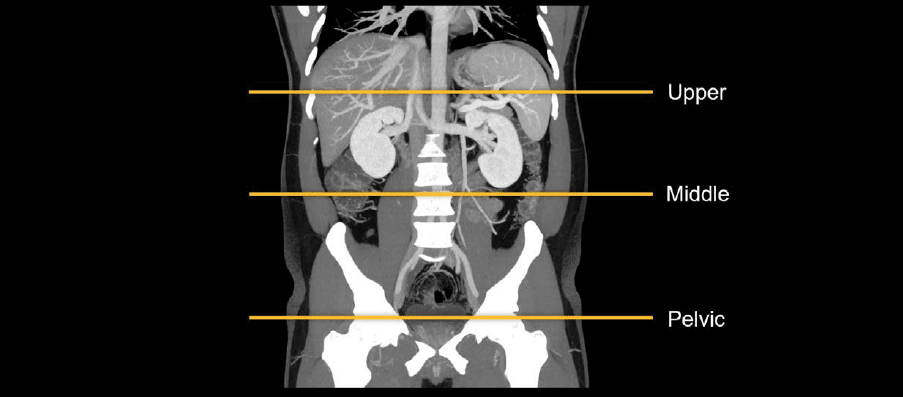
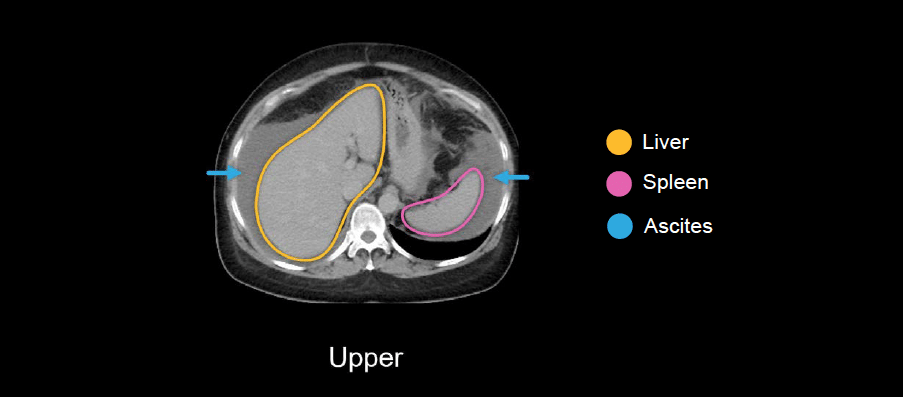
Upper
The liver and spleen are commonly surrounded by ascites when fluid is in the upper abdomen. Ascites will appear darker than the organs but brighter than the fat in the body wall or peritoneal cavity.
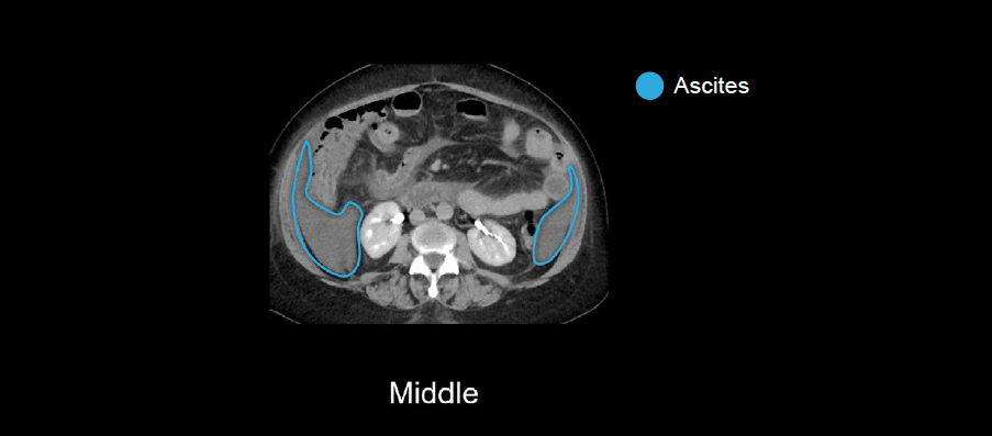
Middle
In the middle abdomen, ascites helps you to see the boundaries of the peritoneal and retroperitoneal cavities and (often) you can also see layers in the sides of the peritoneal cavity near the ascending and descending colon, which are called the paracolic gutter. This is the route where fluid easily moves between the upper abdomen and pelvis.
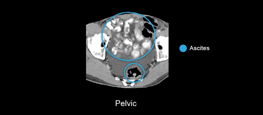
Pelvis
In the pelvis, ascites will surround the small bowel loops and the sigmoid colon and rectum.
Knowledge iteration
To solidify the core anatomy, scroll through a set of images on a PACS viewer. Click on the image below to scroll through a set of images reviewing the peritoneum, omentum, and retroperitoneum.
- Notice that the fat within each of the compartments blends together when they are normal, so you need to use other landmarks to help guide you. You can identify the retroperitoneal space by finding the adrenals, kidneys, pancreas, and ascending and descending colon.
- The peritoneal cavity contains the small bowel, mesentery, and omentum. The omentum is the fatty layer draping over the small bowel.
- You can usually see some vessels running through the omentum, and it will vary in thickness between patients.
This is an edited excerpt from the Medmastery course Abdominal CT Essentials by Michael P. Hartung, MD. Acknowledgement and attribution to Medmastery for providing course transcripts.
- Hartung MP. Abdominal CT: Common Pathologies. Medmastery
- Hartung MP. Abdominal CT: Essentials. Medmastery
- Hartung MP. Abdomen CT: Trauma. Medmastery
References
- Top 100 CT scan quiz. LITFL
Radiology Library: Abdominal CT: Imaging the abdominal organs and bowel
- Hartung MP. Abdominal CT: Evaluating the liver
- Hartung MP. Abdominal CT: Evaluating the biliary system and pancreas
- Hartung MP. Abdominal CT: Imaging the spleen and adrenal glands
- Hartung MP. Abdominal CT: Mastering the genitourinary system
- Hartung MP. Abdominal CT: Evaluating the lower oesophagus and stomach
- Hartung MP. Abdominal CT: Imaging the small bowel
- Hartung MP. Abdominal CT: Viewing the peritoneal cavity
- Hartung MP. Abdominal CT: Running the large bowel and appendix
Abdominal CT interpretation

Assistant Professor of Abdominal Imaging and Intervention at the University of Wisconsin Madison School of Medicine and Public Health. Interests include resident and medical student education, incorporating the latest technology for teaching radiology. I am also active as a volunteer teleradiologist for hospitals in Peru and Kenya. | Medmastery | Radiopaedia | Website | Twitter | LinkedIn | Scopus

MBChB (hons), BMedSci - University of Edinburgh. Living the good life in emergency medicine down under. Interested in medical imaging and physiology. Love hiking, cycling and the great outdoors.