
![]()
Abdominal CT: oesophagus and stomach
Evaluating the lower oesophagus and stomach
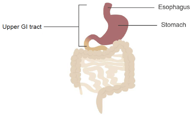
Distal oesophagus
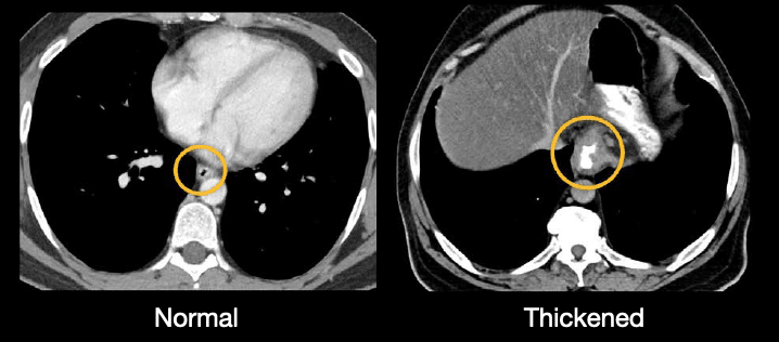
The distal oesophagus is routinely included in an abdominal CT scan (the portion that covers the lower chest). It usually appears as a small, decompressed tube, but you might encounter thickening which can be related to inflammation or a mass.
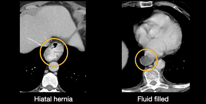
If there is reflux or obstruction, the esophagus may fill with fluid. You may also see a hiatal hernia, where a portion of the stomach has extended above the diaphragm, as shown below. The bright material in the lumen, in this case, is oral contrast.
Stomach
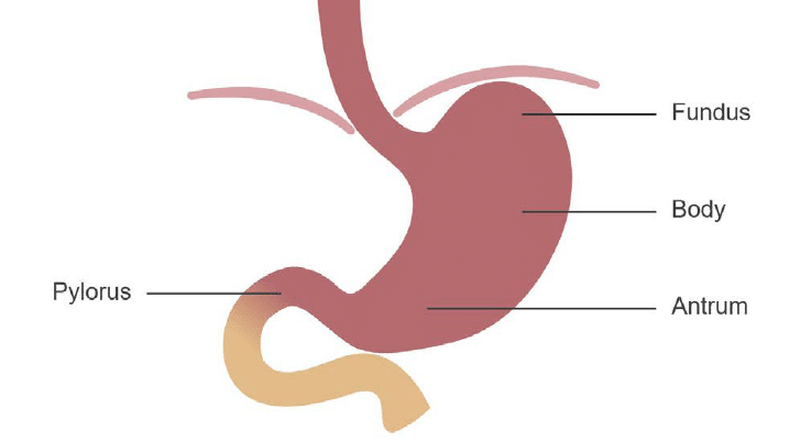
As we follow the distal oesophagus below the diaphragm, we enter the stomach, which is in the peritoneal cavity. The stomach anatomy includes the following four structures:
- Fundus
- Body
- Antrum
- Pylorus
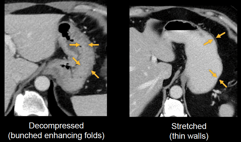
The stomach can vary in appearance depending on when the patient last ate or drank. The stomach can be collapsed when fasting or stretch quite a bit when full. This change will affect your ability to see the typical wrinkly fold pattern which is referred to as rugae.
The example below on the left, shows the typical appearance of a decompressed (i.e., collapsed) stomach where the enhancing folds bunch up. The example on the right shows oral contrast filling the stomach and stretching out the folds, resulting in a thin appearance of the stomach wall.
At the end of the stomach, you will find the pylorus. It can often be seen on CT, and it can have a range of normal thicknesses.
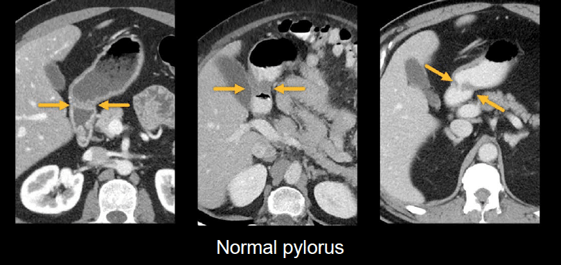
However, if the pylorus looks particularly thickened or is surrounded by fluid and fat stranding, you might become concerned about peptic ulcer disease or a mass.
Examples:
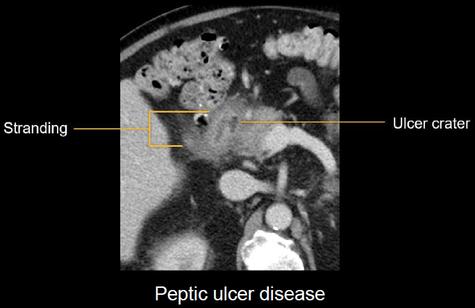
In the first image below, there is an oval ulcer crater with stranding, indicating inflammation of the pylorus wall and surrounding fat due to peptic ulcer disease.
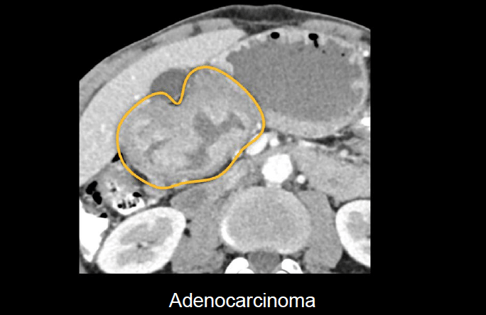
Below, we see a large, irregular mass of the pylorus that was confirmed to be adenocarcinoma after endoscopic biopsy.
This is an edited excerpt from the Medmastery course Abdominal CT Essentials by Michael P. Hartung, MD. Acknowledgement and attribution to Medmastery for providing course transcripts.
- Hartung MP. Abdominal CT: Common Pathologies. Medmastery
- Hartung MP. Abdominal CT: Essentials. Medmastery
- Hartung MP. Abdomen CT: Trauma. Medmastery
References
- Top 100 CT scan quiz. LITFL
Radiology Library: Abdominal CT: Imaging the abdominal organs and bowel
- Hartung MP. Abdominal CT: Evaluating the liver
- Hartung MP. Abdominal CT: Evaluating the biliary system and pancreas
- Hartung MP. Abdominal CT: Imaging the spleen and adrenal glands
- Hartung MP. Abdominal CT: Mastering the genitourinary system
- Hartung MP. Abdominal CT: Evaluating the lower oesophagus and stomach
- Hartung MP. Abdominal CT: Imaging the small bowel
- Hartung MP. Abdominal CT: Viewing the peritoneal cavity
- Hartung MP. Abdominal CT: Running the large bowel and appendix
Abdominal CT interpretation

Assistant Professor of Abdominal Imaging and Intervention at the University of Wisconsin Madison School of Medicine and Public Health. Interests include resident and medical student education, incorporating the latest technology for teaching radiology. I am also active as a volunteer teleradiologist for hospitals in Peru and Kenya. | Medmastery | Radiopaedia | Website | Twitter | LinkedIn | Scopus

MBChB (hons), BMedSci - University of Edinburgh. Living the good life in emergency medicine down under. Interested in medical imaging and physiology. Love hiking, cycling and the great outdoors.

As a resident, very much appreciate this radiology series