![]()
Abdominal CT: small intestine
Imaging the small bowel
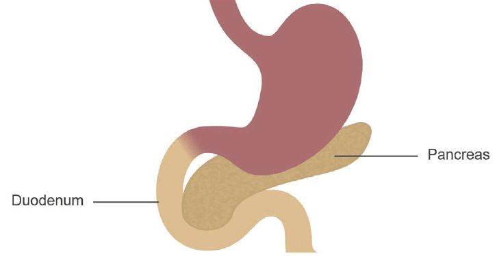
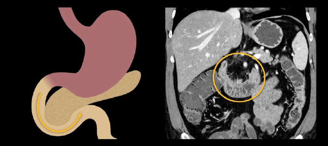
Duodenum
The duodenum is characterized by its c-shape curving around the pancreas
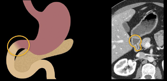
The first portion of the duodenum is the duodenal bulb, which you might recognize on some CT scans by its rounded, light bulb-shape when it is filled with fluid.
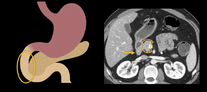
The second portion of the duodenum transitions into the retroperitoneum and runs along the outside of the pancreas head.
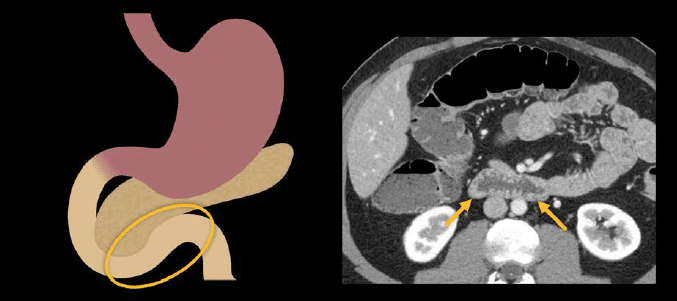
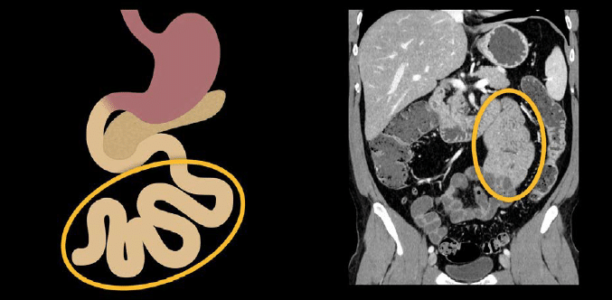
The third portion crosses the midline to the left, heading back into the peritoneal cavity and on to the second section of the small intestine, called the jejunum.
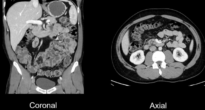
In the image shown next, we can see that C-shape on a coronal image. The sweep-like curve of the duodenum becomes clear in this view as you follow the third portion of the duodenum across the midline toward the jejunum.
Jejunum
The jejunum has a characteristic, feathery, fold pattern and is mostly in the left upper and mid abdomen.
Ileum
The ileum is the third and final section of the small intestine. It has a smoother, simpler-looking wall compared to the jejunum, and it is mostly located in the lower abdomen and right lower quadrant.
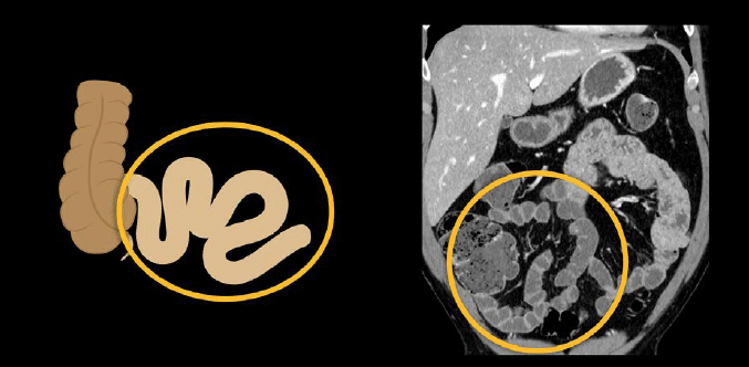
The terminal ileum is the last part of the ileum before you reach the cecum. It is an important site to evaluate for infection and inflammation as it is commonly involved with inflammatory bowel diseases, like Crohn disease.
Evaluating the small intestine for obstruction and inflammation
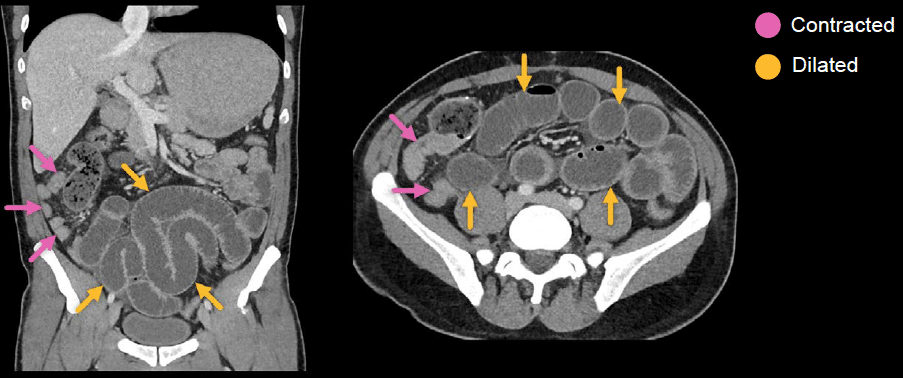
Generally speaking, the small intestine is more intuitive to evaluate on coronal images because you can see so much of it in a single image, and any potentially abnormal segments will stand out compared to the other loops.
However, when you are trying to trace the small intestine, such as in a complex small bowel obstruction, it is usually best to use both coronal and axial views, side-by-side, in the PACS viewer.
CT is excellent for small bowel pathology and is commonly performed to look for obstruction or inflammation. Small bowel obstruction will show dilated (i.e., stretched-out), fluid-filled loops of small bowel that become small in size after the point of obstruction.
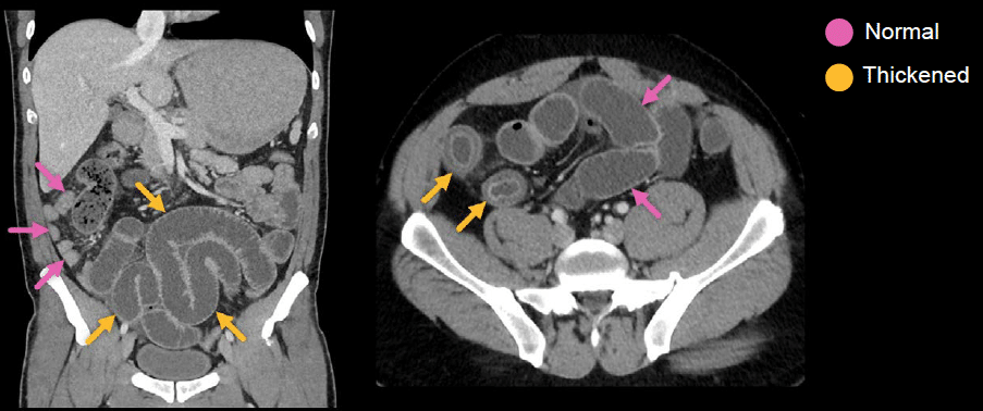
Small bowel wall thickening and abnormal enhancement must be considered in light of the patient’s history and laboratory values, and it can occur from a wide variety of pathologies, including infection, inflammation and ischaemia
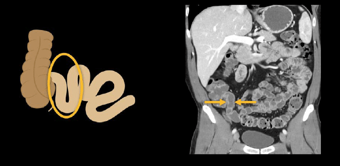
Example: infectious enteritis
Notice how the coronal images help us to detect the abnormally thickened segments of the ileum compared to the normal wall thickness in other loops.
Knowledge iteration
To solidify the core anatomy from the last two lessons, scroll through a set of images on a PACS viewer. click on the image below to scroll through a set of images which show normal appearing upper GI tract and small bowel.
- On the axial images, you can see the distal oesophagus and scroll down into the stomach to review the gastric fundus, body, antrum, and pylorus.
- Note the characteristic shape of the duodenal bulb and scroll down along the second and third portions of the duodenum.
- From there, you can continue your evaluation of the small bowel and notice the fold pattern of the jejunum which transitions to the smoother ileum and, finally, the terminal ileum.
- The coronal images provide a very helpful review as well. Starting with the distal oesophagus, you can see the gastro-oesophageal junction, stomach with characteristic rugal fold pattern, gastric antrum, and pylorus.
- You can then view the second portion of the duodenum sweeping down into the third portion of the duodenum. Next, you can see the jejunum in the left and mid abdomen and follow it to the smooth ileum and finally to the terminal ileum in the right lower quadrant.
- The coronal images make it very easy to look at long segments of small bowel and mesentery.
This is an edited excerpt from the Medmastery course Abdominal CT Essentials by Michael P. Hartung, MD. Acknowledgement and attribution to Medmastery for providing course transcripts.
- Hartung MP. Abdominal CT: Common Pathologies. Medmastery
- Hartung MP. Abdominal CT: Essentials. Medmastery
- Hartung MP. Abdomen CT: Trauma. Medmastery
References
- Top 100 CT scan quiz. LITFL
Radiology Library: Abdominal CT: Imaging the abdominal organs and bowel
- Hartung MP. Abdominal CT: Evaluating the liver
- Hartung MP. Abdominal CT: Evaluating the biliary system and pancreas
- Hartung MP. Abdominal CT: Imaging the spleen and adrenal glands
- Hartung MP. Abdominal CT: Mastering the genitourinary system
- Hartung MP. Abdominal CT: Evaluating the lower oesophagus and stomach
- Hartung MP. Abdominal CT: Imaging the small bowel
- Hartung MP. Abdominal CT: Viewing the peritoneal cavity
- Hartung MP. Abdominal CT: Running the large bowel and appendix
Abdominal CT interpretation

Assistant Professor of Abdominal Imaging and Intervention at the University of Wisconsin Madison School of Medicine and Public Health. Interests include resident and medical student education, incorporating the latest technology for teaching radiology. I am also active as a volunteer teleradiologist for hospitals in Peru and Kenya. | Medmastery | Radiopaedia | Website | Twitter | LinkedIn | Scopus

MBChB (hons), BMedSci - University of Edinburgh. Living the good life in emergency medicine down under. Interested in medical imaging and physiology. Love hiking, cycling and the great outdoors.