![]()
Abdominal CT: Liver
Evaluating the liver
When reviewing the solid organs, use a checklist approach and move systematically through the anatomy. This way, you can make sure to look at all of the important structures and you can reinforce your search pattern.
Liver
The portal venous phase provides the best balance of solid organ and vascular enhancement for general imaging, and it is the most common type of computed tomography (CT) performed
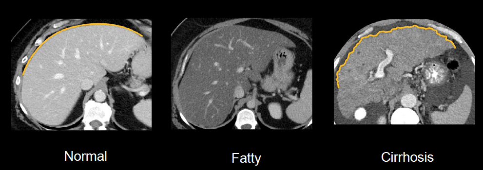
The normal liver tissue has a smooth contour and uniform enhancement. The most common variations that you will encounter are fatty liver and cirrhosis. Fatty liver (i.e., steatosis) will appear less bright than normal liver tissue due to fat infiltration throughout the organ. Cirrhotic livers often have a nodular, bumpy surface.
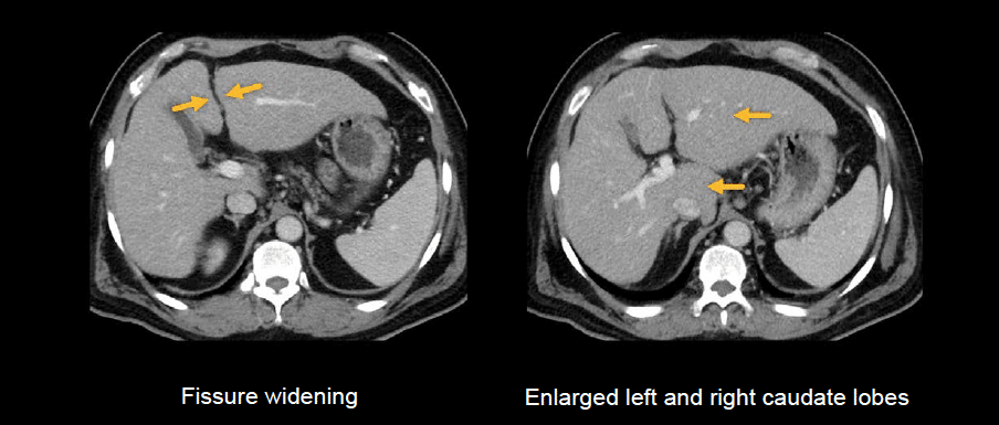
Cirrhosis can also present with more subtle nodularity of the liver surface but will often give other hints of the diagnosis, including widening of the fissure and enlargement of the left and caudate lobes.
When evaluating the liver for different types of masses, there are three types to look for:
- Fluid-filled cyst
- Hemangioma
- Solid masses
Fluid-filled cyst
These benign lesions will appear darker than the surrounding liver tissue and will have a smooth, thin, or imperceptible wall. Patients may have only a few cysts or they can have many, as shown here.
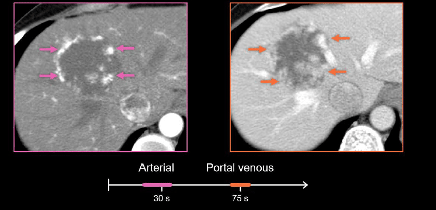
Hemangioma
In contrast to a cyst, a hemangioma has internal enhancement that will fill in over time. These are also benign and relatively common in the liver.
For hemangiomas in the arterial phase, the enhancement starts as small nodular areas along the outside of the mass that grow and fill in toward the middle during the portal venous phase. Seeing this progression helps to confirm the diagnosis.
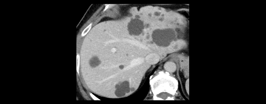
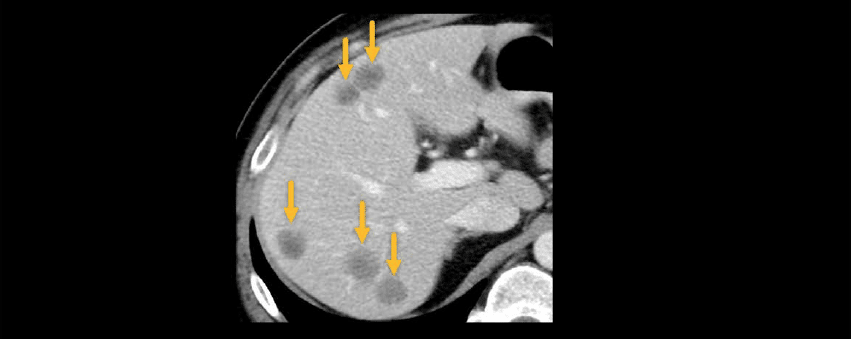
Solid masses
These often enhance less than the liver tissue but do not appear as dark as a cyst. These masses can be benign or malignant. Multiple round masses like the ones shown below are suspicious for metastases and would require further evaluation.
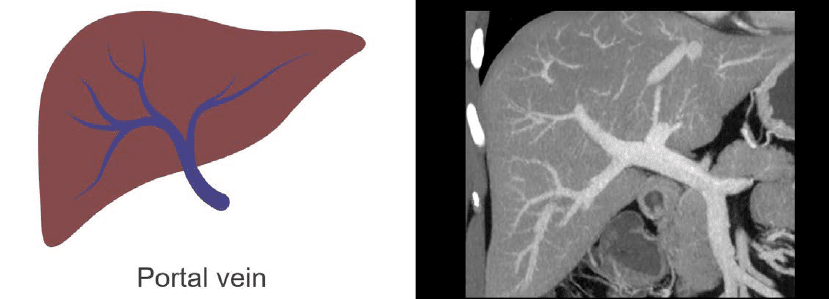
Liver vasculature
When evaluating the liver vasculature, you should first check the portal vein and follow it into the liver as it splits into the right and left branches.
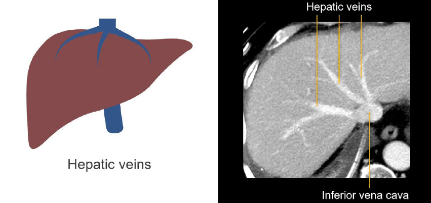
In a portal venous phase CT, the right, middle, and left hepatic veins are usually filled with contrast-mixed blood and can be seen emptying into the inferior vena cava, which is found along the back side of the liver.
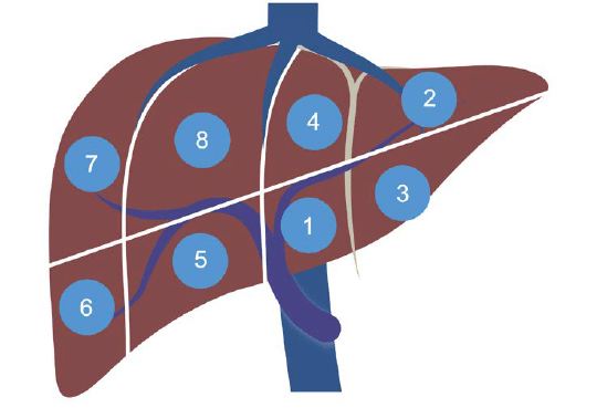
The portal and hepatic veins divide the liver into segmental anatomy that allows radiologists and surgeons to be very specific about the locations of different lesions or abnormalities. The hepatic veins and falciform ligament divide the liver vertically, whereas the portal veins divide the liver horizontally, together forming eight segments.
This is an edited excerpt from the Medmastery course Abdominal CT Essentials by Michael P. Hartung, MD. Acknowledgement and attribution to Medmastery for providing course transcripts.
- Hartung MP. Abdominal CT: Common Pathologies. Medmastery
- Hartung MP. Abdominal CT: Essentials. Medmastery
- Hartung MP. Abdomen CT: Trauma. Medmastery
References
- Top 100 CT scan quiz. LITFL
Radiology Library: Abdominal CT: Imaging the abdominal organs and bowel
- Hartung MP. Abdominal CT: Evaluating the liver
- Hartung MP. Abdominal CT: Evaluating the biliary system and pancreas
- Hartung MP. Abdominal CT: Imaging the spleen and adrenal glands
- Hartung MP. Abdominal CT: Mastering the genitourinary system
- Hartung MP. Abdominal CT: Evaluating the lower oesophagus and stomach
- Hartung MP. Abdominal CT: Imaging the small bowel
- Hartung MP. Abdominal CT: Viewing the peritoneal cavity
- Hartung MP. Abdominal CT: Running the large bowel and appendix
Abdominal CT interpretation

Assistant Professor of Abdominal Imaging and Intervention at the University of Wisconsin Madison School of Medicine and Public Health. Interests include resident and medical student education, incorporating the latest technology for teaching radiology. I am also active as a volunteer teleradiologist for hospitals in Peru and Kenya. | Medmastery | Radiopaedia | Website | Twitter | LinkedIn | Scopus

MBChB (hons), BMedSci - University of Edinburgh. Living the good life in emergency medicine down under. Interested in medical imaging and physiology. Love hiking, cycling and the great outdoors.