
![]()
Dressler beat
Description
Dressler beat: Specifically a ‘ventricular fusion beat‘ in the presence of paroxysmal ventricular tachycardia. Typically observed in ECG tracings of wide complex tachycardia such as VT with AV dissociation.
Supraventricular and a ventricular impulses coincide to produce a hybrid complex which is different to the VT complex and the native complex (capture beat)
The term ‘fusion beat‘ was originally used to define any hybrid QRS complex of atria or ventricular origin. Fusion beats (and capture beats) are not ‘diagnostic‘ or ‘pathognomic‘ of VT and can occur in any arrhythmia (including SVT with aberrancy for example)
History of the Dressler beat
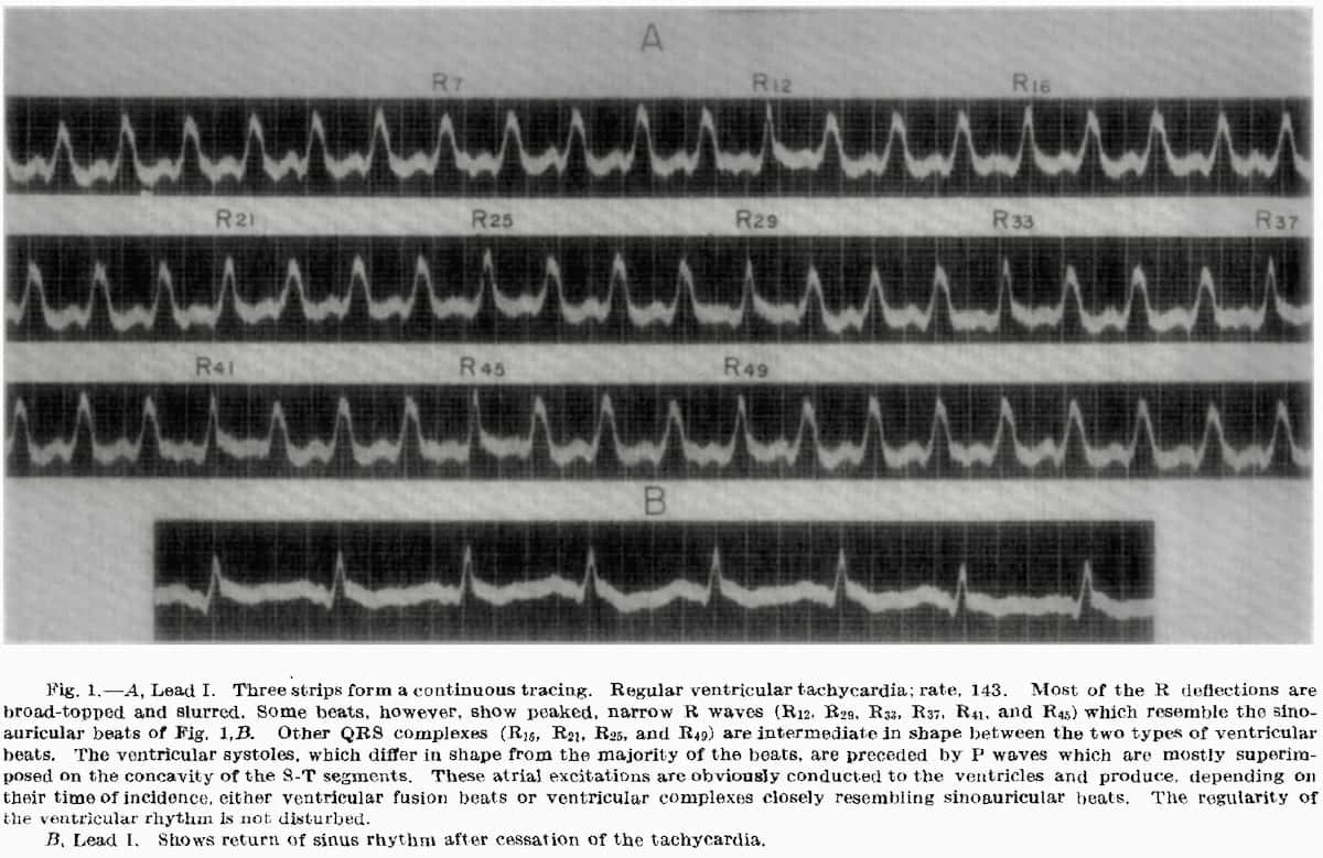
1943 – W. Trevor Cooke and Paul D. White, writing on paroxysmal ventricular tachycardia included two tracings (Fig. 13, Case 21; and Fig. 1, Case 22) which show variations in the shape of the ventricular complexes apparently due to conduction of sinoauricular excitations.
In discussing Fig. 1, Case 22, the authors observe that “…breaks in the ventricular rhythm in Leads I and II are shown…” without commenting on the mechanism involved.
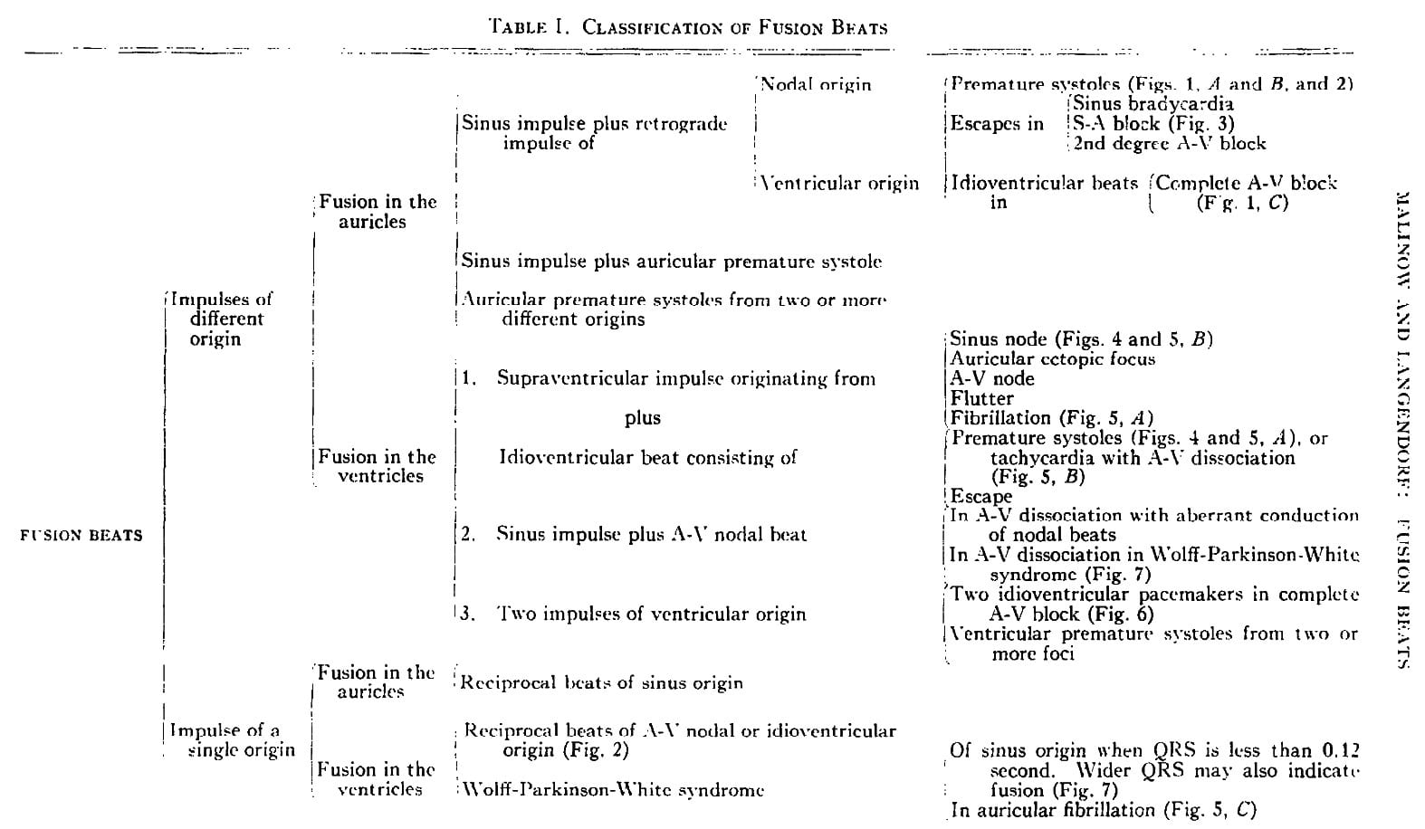
1948 – Malinow and Langendorf developed a classification table for the various types of ‘fusion beat‘ and illustrate the proposed mechanisms for fusion beat formation. As detailed in their proposed classification – fusion beats are not synonymous with VT.
“Since the fusing impulses can be of either different or identical origin, and since fusion which is reflected in the electrogardiagram can take place either in the auricle or in the ventricle four different types of fusion beats can be distinguished. “
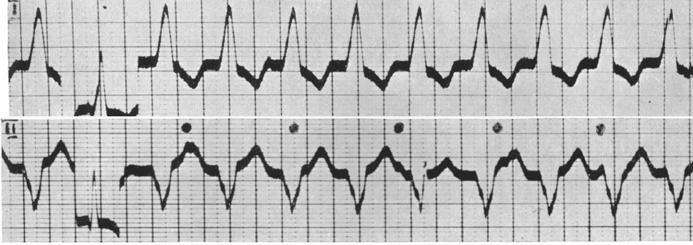
1952 – Dressler stated that “…reports in the literature on the occurrence of ventricular fusion beats in paroxysmal ventricular tachycardia are scant.” He reported six cases of paroxysmal ventricular tachycardia, which demonstrated variations of the ventricular complexes “transitional in shape to sinoauricular beats” and termed them ‘ventricular fusion beats‘.
Six instances of paroxysmal ventricular tachycardia are reported, which show variations of the ventricular complexes transitional in shape to sinoauricular beats. The variations are caused by transmission of atrial excitations to the ventricles, which mostly results in ventricular fusion beats. The regularity of the ventricular rhythm is only exceptionally disturbed by conduction of atrial excitations.
In the presence of paroxysmal ventricular tachycardia, dissociation with interference develops infrequently because of the shortness of the ventricular cycle. When it does occur, the ventricular rate is usually less than 150 per minute
The finding of ventricular complexes which are transitional in configuration to sinoauricular beats may be an aid in the diagnosis of paroxysmal ventricular tachycardia when an independent atrial rhythm is not readily recognizable.
Dressler W: Am Heart J. 1952 ;44(4):485-493
1962 – Marriott, Schwartz and Bix reviewed and demonstrated stages of ventricular fusion beat development in a multitude of rhythms. They define ventricular fusion as a
…summation or combination beats occur when two separate pacemakers compete for control of the ventricles. The usual pair of pacemakers are the sinoatrial node and an ectopic ventricular focus; but any pair of pacemakers, whose impulses invade the ventricular myocardium more or less simultaneously but at different points, can produce fusion beats.
Marriott: Circulation. 1962
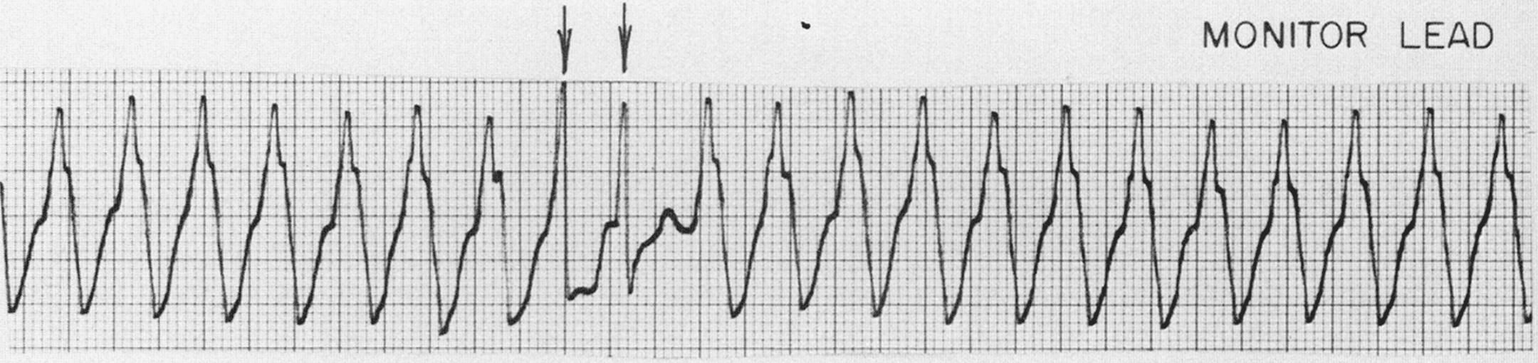
Young et al Dressler beats in VT (1973)

(arrows). Third beat marked is especially narrow and represents almost complete supraventricular capture. [Young et al: Chest. 1973]
Controversies
Potentially we should revert to the use of ‘Dressler beat‘ in the presence of VT. The term ‘fusion beat’ was originally used to define any hybrid QRS complex of atria or ventricular origin. Fusion beats (and capture beats) are not ‘diagnostic‘ of VT and can occur in any arrhythmia (including SVT with aberrancy for example)
Various publications cite Dressler beat as either fusion beat or capture beat. Dressler describes the abnormal hybrid complex associated with ventricular fusion beats (Dressler beat) as opposed to a native complex in VT (capture beat)
During four episodes of ventricular tachycardia, capture beats (Dressler beats) led to an irregular ventricular rhythm.
Wellens (1978)
The conducted supraventricular beat may arrive coincident with discharge of the ectopic focus, in which case complexes may emerge with configurations intermediate between normal and those of the VT.
Lown (1973)
Associated Persons
- Paul Dudley White (1886–1973)
- Louis Nelson Katz (1897 – 1973)
- Wilhelm Dressler (1890–1969)
- Bernard Lown (1921 – )
- Hein Wellens (1935 – )
Alternative names
- Fusion beat
- Ventricular fusion beat
References
- Cooke WT, White PD. Paroxysmal ventricular tachycardia. Br Heart J. 1943 Jan; 5(1): 33–54
- Katz LN. Electrocardiography. Lea & Febiger, Philadelphia. 1947 [Figure 322, 328]
- Malinow MR, Langendorf R. Different mechanisms of fusion beats. Am Heart J. 1948;35(3):448-457
- Malinow-Langendorf. Proposed Classification of Fusion Beats, 1948
- Dressler W, Roesler H. The occurrence in paroxysmal ventricular tachycardia of ventricular complexes transitional in shape to sinoauricular beats: A diagnostic aid. Am Heart J. 1952 Oct;44(4):485-93.
- Marriott HJL, Schwartz NL, Bix HH. Ventricular Fusion Beats. Circulation. 1962;26:880-884
- Young RL, Mower MM, Ramapuram GM, Tabatznik B. Atrial fibrillation with ventricular tachycardia showing “Dressler” beats. Chest. 1973 Jan;63(1):96-97
- Lown B, Temte JV, Arter WJ. Ventricular Tachyarrhythmias: Clinical aspects. Circulation. 1973;47:1364-1381
- Wellens HJ, Bär FW, Lie KI. The value of the electrocardiogram in the differential diagnosis of a tachycardia with a widened QRS complex. Am J Med. 1978 Jan;64(1):27-33
- Smith S. Wide Complex Tachycardia with Fusion and Capture Beats. Not what you think. Dr. Smith’s ECG Blog. 2016
- Burns E. Fusion Beats. LITFL. 2017
LITFL Further Reading
- ECG Library Basics – Waves, Intervals, Segments and Clinical Interpretation
- ECG A to Z by diagnosis – ECG interpretation in clinical context
- ECG Exigency and Cardiovascular Curveball – ECG Clinical Cases
- 100 ECG Quiz – Self-assessment tool for examination practice
- ECG Reference SITES and BOOKS – the best of the rest
Advanced Reading
- Brady WJ, Truwit JD. Critical Decisions in Emergency and Acute Care Electrocardiography
- Surawicz B, Knilans T. Chou’s Electrocardiography in Clinical Practice: Adult and Pediatric
- Wagner GS. Marriott’s Practical Electrocardiography 12e
- Chan TC. ECG in Emergency Medicine and Acute Care
- Rawshani A. Clinical ECG Interpretation
- Mattu A. ECG’s for the Emergency Physician
- Hampton JR. The ECG In Practice, 6e
ECG LIBRARY
BA MA (Oxon) MBChB (Edin) FACEM FFSEM. Emergency physician, Sir Charles Gairdner Hospital. Passion for rugby; medical history; medical education; and asynchronous learning #FOAMed evangelist. Co-founder and CTO of Life in the Fast lane | On Call: Principles and Protocol 4e| Eponyms | Books |

MBBS FACEM DDU (Emergency) CCPU. Emergency Physician in Melbourne, Australia. Co-Ultrasound Lead for Emergency Medicine at The Alfred Hospital. Special interests in diagnostic and procedural ultrasound, medical education, and ECG interpretation. Editor of the LITFL ECG Library.
