ECG Case 037
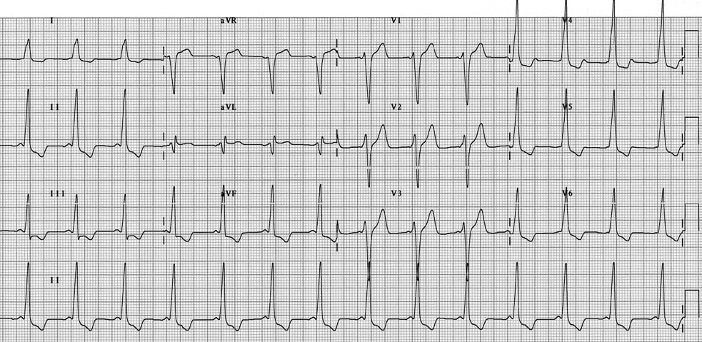
15-year old patient presenting with rapid palpitations and dizziness. Symptoms resolved en route to hospital. Describe the ECG.

Describe and interpret this ECG
ECG ANSWER and INTERPRETATION
Main Abnormalities
This ECG is diagnostic of the Wolff-Parkinson-White (WPW) syndrome:
- Sinus rhythm with a very short PR interval (< 120ms)
- Broad QRS complexes
- Delta waves = slurred upstroke to the QRS
Other Features:
- Dominant S wave in V1 — this “type B” pattern indicates a right-sided accessory pathway
- Tall R waves and inverted T waves mimic the appearance of LVH — this is an electrical phenomenon due to WPW and not a sign of ventricular hypertrophy
- ST segments and T waves show typical “discordant” changes — they point in in the opposite direction to the QRS complex, similar to LBBB
References
Further Reading
- Wiesbauer F, Kühn P. ECG Mastery: Yellow Belt online course. Understand ECG basics. Medmastery
- Wiesbauer F, Kühn P. ECG Mastery: Blue Belt online course: Become an ECG expert. Medmastery
- Kühn P, Houghton A. ECG Mastery: Black Belt Workshop. Advanced ECG interpretation. Medmastery
- Rawshani A. Clinical ECG Interpretation ECG Waves
- Smith SW. Dr Smith’s ECG blog.
- Wiesbauer F. Little Black Book of ECG Secrets. Medmastery PDF
TOP 100 ECG Series
Emergency Physician in Prehospital and Retrieval Medicine in Sydney, Australia. He has a passion for ECG interpretation and medical education | ECG Library |
MBBS DDU (Emergency) CCPU. Adult/Paediatric Emergency Medicine Advanced Trainee in Melbourne, Australia. Special interests in diagnostic and procedural ultrasound, medical education, and ECG interpretation. Co-creator of the LITFL ECG Library. Twitter: @rob_buttner

