


Reimers migration percentage
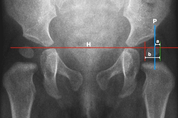
Reimers migration percentage (MP) quantifies lateral migration of the visible femoral head on an AP pelvic radiograph.
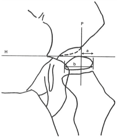
In modern practice, Perkins line is drawn perpendicular to the pelvic horizontal through the lateral edge of the acetabular roof. The horizontal width of the visible femoral head lateral to Perkins line (a) is divided by the total horizontal width of the visible femoral head (b):
MP = a / b × 100
Hilgenreiner line is used to establish the pelvic horizontal from which Perkins line is constructed, but the horizontal line used to measure a and b is a separate measurement through the femoral head.
Original Reimers method
Reimers chose Perkins line because it provided a direct measure of the position of the femoral head relative to the acetabulum and avoided the difficulty of locating the centre of the femoral head required for the centre-edge angle.
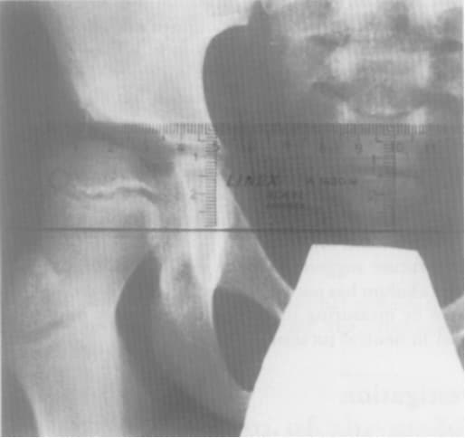
In his 1980 study, Reimers defined subluxation as MP ≥33% and dislocation as MP 100%. These were study definitions and should be distinguished from thresholds used in later hip-surveillance programmes.
MP = a / b × 100
Historical development of the construction
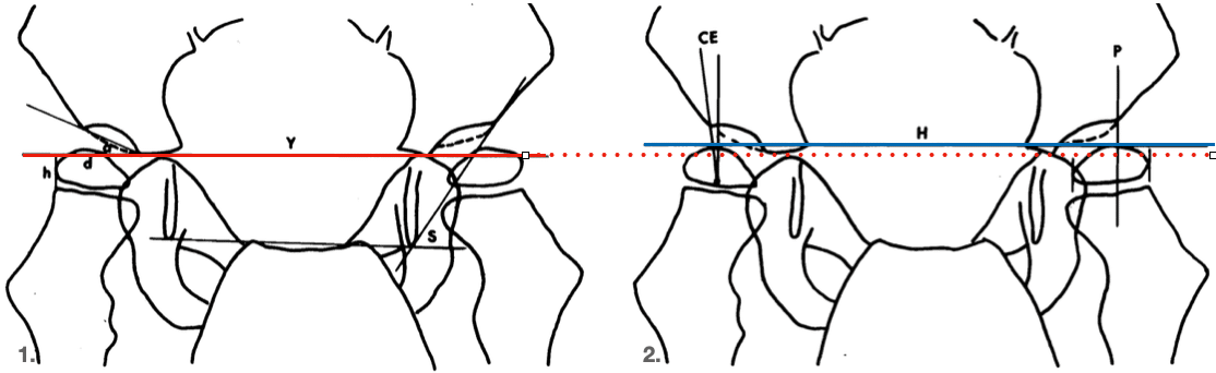
Reimers’ historical review distinguished two related constructions. In his Figure 1, the Y-line is identified as the Hilgenreiner line and passes through the triradiate cartilages. In Figure 2, the horizontal reference used to construct Perkins line is labelled simply H and is drawn through the innermost bony portions of the ilia at the level of the Y-cartilages.
Hilgenreiner defined the line only as connecting the two triradiate cartilages, allowing variation in the precise radiographic landmark used by later authors. Reimers did not specifically identify H in Figure 2 as Hilgenreiner line, and his drawing demonstrates a more caudal position of the line H compared to Y. Later authors combined the two named lines as the Hilgenreiner–Perkins construction.
History
1925 – Heinrich Hilgenreiner described a horizontal baseline connecting the two triradiate cartilages as part of his radiographic system for congenital hip dislocation.
1928 – George Perkins described his perpendicular acetabular reference line (Perkins line). Reimers characterised it as running from the lateral acetabular region perpendicular to a horizontal reference at the Y-cartilages.
1950 – Heyman and Herndon described an acetabulum–head index based on femoral-head coverage.
1961 – Medbø measured in millimetres the portion of the femoral head projecting lateral to Perkins line. Zweymüller and Wicke used a similar measurement in 1973.
1975 – Mercer Rang suggested the term migration percentage to Reimers.
1979–1980 – Jørgen Reimers formalised the measurement as migration percentage (MP), investigated positioning, and established normal and pathological values. Reimers applied serial MP to the study of hip migration and muscle surgery in cerebral palsy.
2004 – Faraj et al studied reproducibility of MP and illustrated the later H, P, and a/b construction.
Reimers used the lateral edge of the acetabular roof as the landmark for Perkins line. Later authors proposed a modified technique using the lateral edge of the acetabular sourcil. Subsequent comparative studies have found greater inter-rater reliability with the original acetabular-edge method.
2015 – Analan and colleagues compared migration-percentage measurements made by two physicians with different levels of experience. They found excellent intra- and inter-rater reliability, with inter-rater correlations of 0.94. They concluded that experience level did not materially affect MP measurement when standardised radiographs and a defined technique were used. Their used Hilgenreiner line between the Y-cartilages and Perkins line through the lateral acetabular margin.
2023 – Amen et al. validated smartphone-based measurement of Reimers MP using the HipScreen app across an interdisciplinary clinical team. The study also highlighted a modern variation in the construction. Som methods place the vertical reference at the lateral edge of the sourcil rather than the lateral acetabular edge. The authors retained the classic acetabular-edge landmark as it had demonstrated greater reliability.
Eponymythology
Migration percentage or migration index?
Reimers used migration percentage (MP) for the proportion of the femoral head lateral to Perkins line at a given examination. He used migration index (MI) for the rate of change of MP over time, expressed per year.
Later literature increasingly used migration index as a synonym for migration percentage. Thus, the commonly encountered term “Reimers migration index” does not reproduce Reimers’ original terminology precisely.
New measurement built on old ideas
Reimers did not originate the underlying pelvic lines or the general concept of femoral-head migration. His contribution was to turn lateral displacement into a simple percentage. He defined the measurement technique and applied it longitudinally to the dynamic hip in cerebral palsy.
Which horizontal line?
Reimers’ own figures distinguish Hilgenreiner’s historical Y-line from the horizontal reference used to illustrate Perkins line. Later literature has merged these anatomically different constructions into the standard modern Hilgenreiner–Perkins system.
Associated Persons
- Heinrich Hilgenreiner (1870-1953) and the Hilgenreiner line
- George Perkins (1892-1979) and the Perkins line
- Jørgen Reimers (1931-2008)
Alternative names
- Reimers migration percentage, Migration percentage (MP)
- Reimers hip migration percentage, Hip migration percentage
- Reimers migration index / hip migration index: widely used modern synonym, but historically Reimers used migration index for the annual rate of change.
- Extrusion index: geometrically related and sometimes treated as synonymous, particularly in adult hip radiology.
References
Historical references
- Mezzari A. Sul valore del segno di Shenton per la diagnosi radiografica della lussazione congenita dell’anca nella prima infanzia. La Radiologia Medica, 1928; 15: 13
- Heyman CH, Herndon CH. Legg-Perthes disease; a method for the measurement of the roentgenographic result. J Bone Joint Surg Am. 1950 Oct;32 A(4):767-78.
- Mathews SS, Jones MH, Sperling SC. Hip derangements seen in cerebral palsied children. American Journal of Physical Medicine. 1953 Aug;32(4):213-21.
- Medbø IU. Early diagnosis and treatment of hip joint dysplasia. Acta Orthop Scand. 1961;31:282-315.
- Zweymüller K, Wicke B. Zur Lateralisation des Oberschenkelkopfes beim M. Perthes [Lateralisation of the femoral head in Perthes’ disease]. Arch Orthop Unfallchir. 1973 Apr 24;75(3):239-48.
- Reimers J. The stability of the hip in children. A radiological study of the results of muscle surgery in cerebral palsy. Acta Orthop Scand Suppl. 1980;184:1-100.
Eponymous term review
- Faraj S, Atherton WG, Stott NS. Inter- and intra-measurer error in the measurement of Reimers’ hip migration percentage. J Bone Joint Surg Br. 2004 Apr;86(3):434-7.
- Analan PD, Yilmaz EE, Adam M, Leblebici B. The effect of physician experience on the measurement reliability of the Reimers’ hip migration percentage in children with cerebral palsy. J Phys Ther Sci. 2015 Oct;27(10):3255-8.
- Amen J, Perkins O, Cadwgan J, Cooke SJ, Kafchitsas K, Kokkinakis M. Measuring hip migration percentage in cerebral palsy using the HipScreen app. Bone Jt Open. 2023 May 22;4(5):363-369.
- Kyle E, Cadogan M. Hip and pelvic lines and arcs. LITFL
eponymictionary
the names behind the name

MB BAO BCh Trinity College Dublin, MSc Clinical Data Analytics University of Galway. Junior doctor in Perth, doctoral researcher in MRI radiomics. Working towards radiology training.

BA MA (Oxon) MBChB (Edin) FACEM FFSEM. Emergency physician, Sir Charles Gairdner Hospital. Passion for rugby, medical history, medical education, and asynchronous learning #FOAMed evangelist. Editor-in-Chief and Webmaster of Life in the Fast lane, LITFL | On Call 4e| Eponyms | Books | Horology |