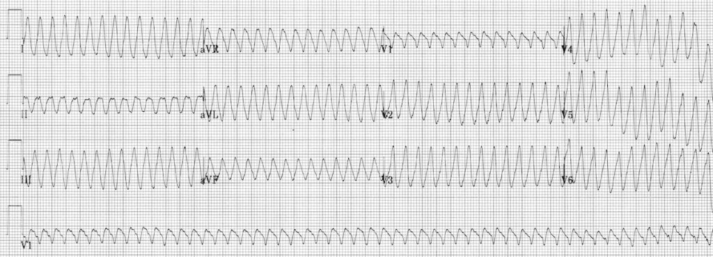
Ventricular Flutter
Clinical Significance of Ventricular flutter
Extreme form of ventricular tachycardia (VT) with loss of organised electrical activity
- Associated with rapid and profound hemodynamic compromise
- Usually short lived due to progression to ventricular fibrillation
- As with ventricular fibrillation rapid initiation of advanced life support is required
How to Recognise Ventricular Flutter
- Continuous Sine Wave
- No identifiable P waves, QRS complexes, or T waves
- Rate usually > 200 beats / min
Clinical Pearl
The ECG looks identical when viewed upside down!
ECG Examples
Example 1

Typical appearance of ventricular flutter:
- Monomorphic sine wave at >200bpm.
- ECG looks identical when turned upside down.
Example 2

12-lead ECG example of ventricular flutter:
- Extremely rapid monomorphic sine wave at around 300 bpm.
Example 3
Ventricular flutter following a bolus of intravenous verapamil

- A supraventricular tachycardia converts to ventricular flutter after administration of verapamil. The rhythm subsequently degenerates into ventricular fibrillation.
- The rapid deterioration with verapamil suggests that the patient may have underlying Wolff-Parkinson White syndrome.
- In WPW, administration of verapamil or diltiazem during a supraventricular tachycardia may produce a paradoxical increase in ventricular rate by increasing conduction through the accessory pathway. With rapid atrial rhythms such as AF or flutter, the sudden onset of 1:1 AV conduction may produce ventricular rates of >300 beats per minute (i.e. ventricular flutter), which rapidly deteriorates to VF.
Related Topics
Advanced Reading
Online
- Wiesbauer F, Kühn P. ECG Mastery: Yellow Belt online course. Understand ECG basics. Medmastery
- Wiesbauer F, Kühn P. ECG Mastery: Blue Belt online course: Become an ECG expert. Medmastery
- Kühn P, Houghton A. ECG Mastery: Black Belt Workshop. Advanced ECG interpretation. Medmastery
- Rawshani A. Clinical ECG Interpretation ECG Waves
- Smith SW. Dr Smith’s ECG blog.
- Wiesbauer F. Little Black Book of ECG Secrets. Medmastery PDF
Textbooks
- Zimmerman FH. ECG Core Curriculum. 2023
- Mattu A, Berberian J, Brady WJ. Emergency ECGs: Case-Based Review and Interpretations, 2022
- Straus DG, Schocken DD. Marriott’s Practical Electrocardiography 13e, 2021
- Brady WJ, Lipinski MJ et al. Electrocardiogram in Clinical Medicine. 1e, 2020
- Mattu A, Tabas JA, Brady WJ. Electrocardiography in Emergency, Acute, and Critical Care. 2e, 2019
- Hampton J, Adlam D. The ECG Made Practical 7e, 2019
- Kühn P, Lang C, Wiesbauer F. ECG Mastery: The Simplest Way to Learn the ECG. 2015
- Grauer K. ECG Pocket Brain (Expanded) 6e, 2014
- Surawicz B, Knilans T. Chou’s Electrocardiography in Clinical Practice: Adult and Pediatric 6e, 2008
- Chan TC. ECG in Emergency Medicine and Acute Care 1e, 2004
LITFL Further Reading
- ECG Library Basics – Waves, Intervals, Segments and Clinical Interpretation
- ECG A to Z by diagnosis – ECG interpretation in clinical context
- ECG Exigency and Cardiovascular Curveball – ECG Clinical Cases
- 100 ECG Quiz – Self-assessment tool for examination practice
- ECG Reference SITES and BOOKS – the best of the rest
ECG LIBRARY
Emergency Physician in Prehospital and Retrieval Medicine in Sydney, Australia. He has a passion for ECG interpretation and medical education | ECG Library |
MBBS DDU (Emergency) CCPU. Adult/Paediatric Emergency Medicine Advanced Trainee in Melbourne, Australia. Special interests in diagnostic and procedural ultrasound, medical education, and ECG interpretation. Co-creator of the LITFL ECG Library. Twitter: @rob_buttner

