![]()
Apical hypertrophic cardiomyopathy (AHC)
ECG features of AHC
- The classic ECG finding in apical HCM is giant T-wave inversion in the precordial leads. Inverted T waves are also commonly observed in the inferior and lateral leads
- Localised hypertrophy of LV apex, causing an “ace of spades” configuration of the LV cavity on ventriculography
Non-obstructive variant of Hypertrophic Cardiomyopathy (HCM). This relatively uncommon form of HCM is seen most frequently in Japanese patients (13-25% of all HCM cases in Japan).
Also known as Yamaguchi syndrome after the Hiroshi Yamaguchi MD and his original papers on hypertrophic non-obstructive cardiomyopathy with giant negative T waves (apical hypertrophy) in 1976 and 1979.
1979 Yamaguchi et al reviewed 1,002 consecutive left heart catheterization patients. Of the total, 30 patients exhibited the pathognomonic “ace of spades” shape of the left ventricle (LV).
2003 Kitaoka et al. showed a prevalence of 15% in an unselected Japanese cohort compared with 3% in an unselected Minneapolis cohort. They found an increased incidence among men, confirmed by Husser et al (2018) who found the incidence increased with age and further favoured men as age increased
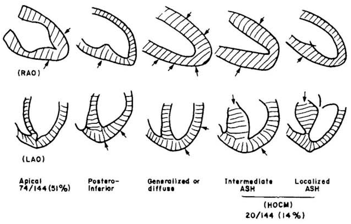
Ace-of-spades sign
Pathognomonic left ventricule configuration seen in apical hypertrophic cardiomyopathy. Marked ventricular wall thickening at the apex resulting in cavity narrowing at the apex with a relatively normal appearance of the mid-ventricular to basal wall and cavity.
The sign can be demonstrated on echocardiography, ventriculography, and cardiac MRI
Ventriculogram
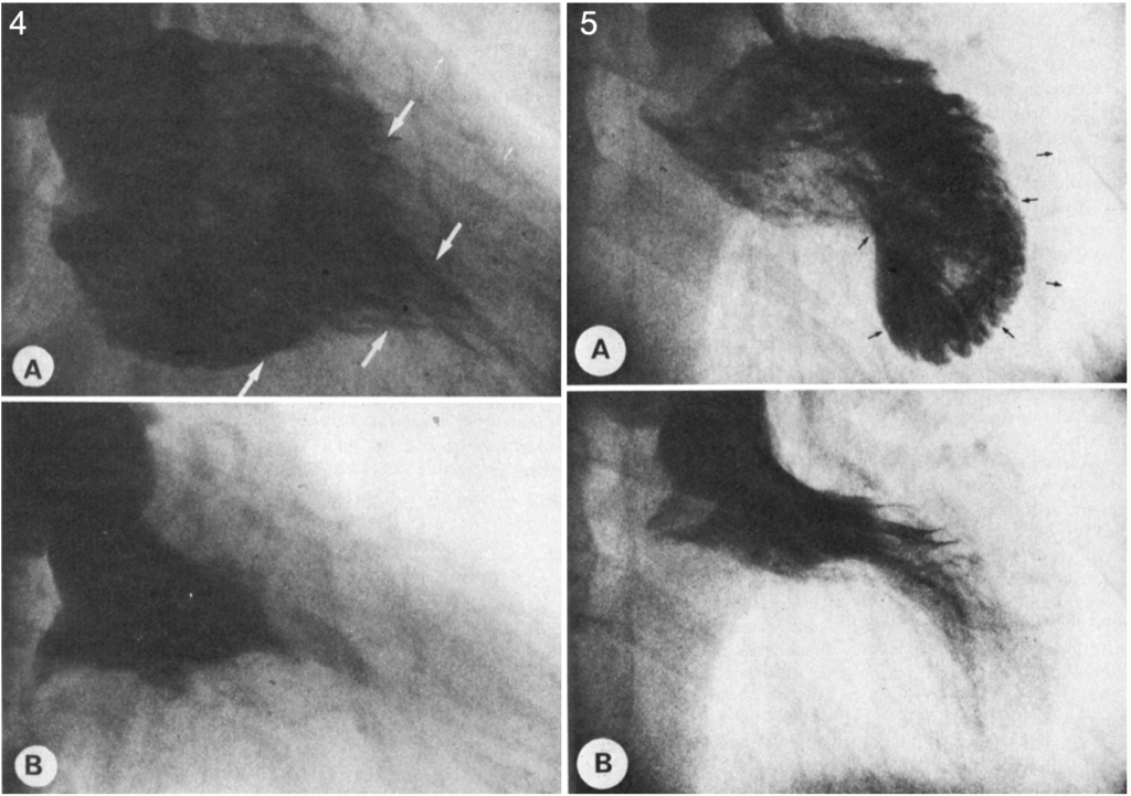
5: Left ventriculograms in hypertrophic obstructive cardiomyopathy at (A) end-diastole the free wall is equally thickened in all segments (arrows) and the left ventricular silhouette is shaped like a kidney or banana. (B), at end systole.
Yamaguchi 1979
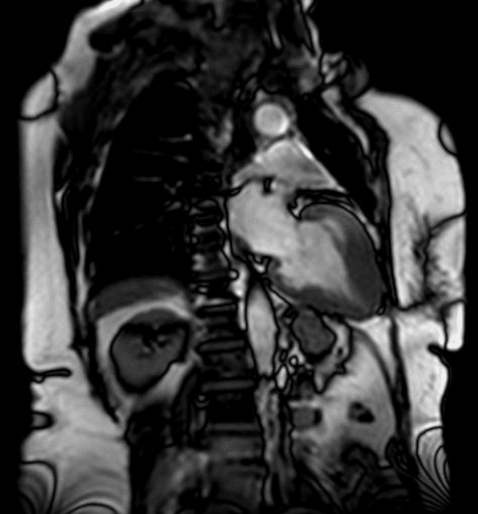
Cardiac MRI
A cardiac MRI with findings suggestive of apical hypertrophic cardiomyopathy. The left ventricle is observed in the right view at the end of diastole with “ace of spades” shape, characteristic of Yamaguchi syndrome
Examples
Example 1
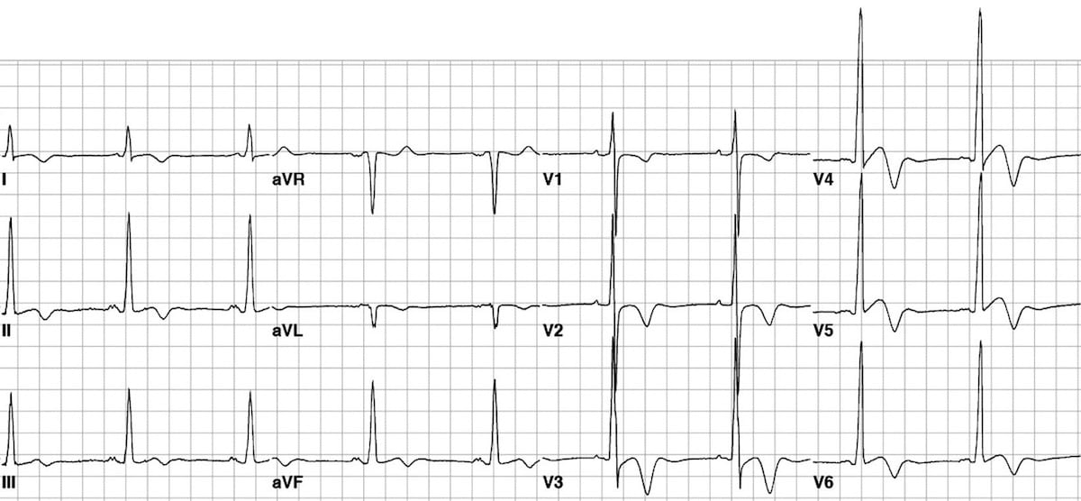
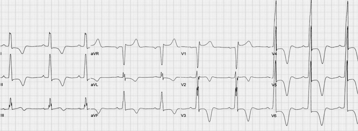
This ECG shows the typical pattern of apical HCM:
- Large precordial voltages.
- Giant T wave inversions in the precordial leads
- Inverted T waves are also seen in the inferior and lateral leads.
This great ECG is reproduced from Hansen & Merchant (2007).
Example 2
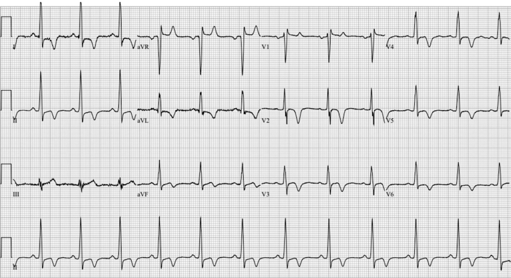
This ECG shows the typical pattern of apical HCM:
- T wave inversions in leads I, II, and V2-V6
Videos
HCM video education
Related Topics
- Dilated cardiomyopathy
- Hypertrophic Cardiomyopathy (HCM)
- Restrictive cardiomyopathy
- Left ventricular hypertrophy
Advanced Reading
References
- Yamagushi H, Nakanishi S, Nishijo T, Nagasaki F, Matsumoto S, Ishimura T. Hypertrophic cardiomyopathy with giant negative T waves. Japanese Circulation Journal. 1976; 40(suppl):110-111.
- Yamaguchi H et al. Hypertrophic nonobstructive cardiomyopathy with giant negative T waves (apical hypertrophy): ventriculographic and echocardiographic features in 30 patients. Am J Cardiol. 1979 Sep;44(3):401-12
- Yamaguchi H, Nishiyama S, Nakanishi S, Nishimura S. Electrocardiographic, echocardiographic and ventriculographic characterization of hypertrophic non-obstructive cardiomyopathy. Eur Heart J. 1983 Nov;4 Suppl F:105-19
- Kitaoka H, Doi Y, Casey SA, Hitomi N, Furuno T, Maron BJ. Comparison of prevalence of apical hypertrophic cardiomyopathy in Japan and the United States. Am J Cardiol. 2003 Nov 15;92(10):1183-6.
- Otieno H, Vivas Y, Traub D, Raman A, Polam C. Images in cardiovascular medicine: contrast echocardiography in apical hypertrophic cardiomyopathy. Circulation. 2006 Jul 11;114(2):e33-4
- van der Wall EE, Bax JJ, Schalij MJ. Detection of apical hypertrophic cardiomyopathy; which is the appropriate imaging modality. Int J Cardiovasc Imaging. 2008 Oct;24(7):683-5.
- Siewe D, Nichols KB, Furney SL, Littmann L. King of hearts for ace of spades: apical hypertrophic cardiomyopathy. Am J Med. 2014 Jan;127(1):31-3.
- Abugroun A, Ahmed F, Vilchez D, Turaga L. Apical Hypertrophic Cardiomyopathy: A Case Report. Cardiol Res. 2017 Oct;8(5):265-268.
- Husser D, Ueberham L, Jacob J, Heuer D, Riedel-Heller S, Walker J, Hindricks G, Bollmann A. Prevalence of clinically apparent hypertrophic cardiomyopathy in Germany-An analysis of over 5 million patients. PLoS One. 2018 May 3;13(5):e0196612.
- Morales J, Giraldo M. Case report: Apical variant hypertrophic cardiomyopathy simulating an acute inferior myocardial infarction. J Electrocardiol. 2019 Jan-Feb;52:35-37
- Shah FA, Fujikawa P, Miller JB, Singh H. A Novel Case of Yamaguchi Syndrome in a Hispanic Male. Cureus. 2021 Sep 1;13(9):e17651.
- Titus A, Sharma N, Narayan G, Sattar Y, Angelis D. From Takotsubo to Yamaguchi. Cureus. 2022 Mar 28;14(3):e23561.
Online
- Wiesbauer F, Kühn P. ECG Mastery: Yellow Belt online course. Understand ECG basics. Medmastery
- Wiesbauer F, Kühn P. ECG Mastery: Blue Belt online course: Become an ECG expert. Medmastery
- Kühn P, Houghton A. ECG Mastery: Black Belt Workshop. Advanced ECG interpretation. Medmastery
- Rawshani A. Clinical ECG Interpretation ECG Waves
- Smith SW. Dr Smith’s ECG blog.
- Wiesbauer F. Little Black Book of ECG Secrets. Medmastery PDF
Textbooks
- Zimmerman FH. ECG Core Curriculum. 2023
- Mattu A, Berberian J, Brady WJ. Emergency ECGs: Case-Based Review and Interpretations, 2022
- Straus DG, Schocken DD. Marriott’s Practical Electrocardiography 13e, 2021
- Brady WJ, Lipinski MJ et al. Electrocardiogram in Clinical Medicine. 1e, 2020
- Mattu A, Tabas JA, Brady WJ. Electrocardiography in Emergency, Acute, and Critical Care. 2e, 2019
- Hampton J, Adlam D. The ECG Made Practical 7e, 2019
- Kühn P, Lang C, Wiesbauer F. ECG Mastery: The Simplest Way to Learn the ECG. 2015
- Grauer K. ECG Pocket Brain (Expanded) 6e, 2014
- Surawicz B, Knilans T. Chou’s Electrocardiography in Clinical Practice: Adult and Pediatric 6e, 2008
- Chan TC. ECG in Emergency Medicine and Acute Care 1e, 2004
LITFL Further Reading
- ECG Library Basics – Waves, Intervals, Segments and Clinical Interpretation
- ECG A to Z by diagnosis – ECG interpretation in clinical context
- ECG Exigency and Cardiovascular Curveball – ECG Clinical Cases
- 100 ECG Quiz – Self-assessment tool for examination practice
- ECG Reference SITES and BOOKS – the best of the rest
ECG LIBRARY

MBBS FACEM DDU (Emergency) CCPU. Emergency Physician in Melbourne, Australia. Co-Ultrasound Lead for Emergency Medicine at The Alfred Hospital. Special interests in diagnostic and procedural ultrasound, medical education, and ECG interpretation. Editor of the LITFL ECG Library.
BA MA (Oxon) MBChB (Edin) FACEM FFSEM. Emergency physician, Sir Charles Gairdner Hospital. Passion for rugby; medical history; medical education; and asynchronous learning #FOAMed evangelist. Co-founder and CTO of Life in the Fast lane | On Call: Principles and Protocol 4e| Eponyms | Books |