![]()
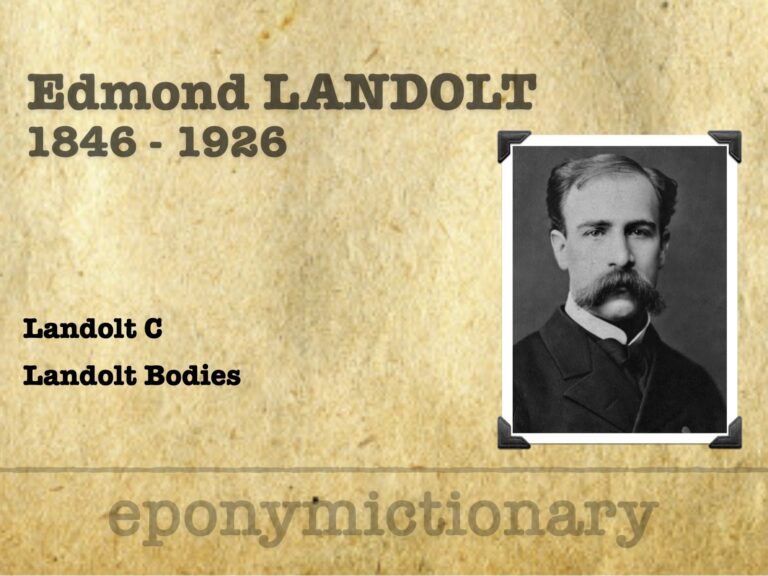
Edmond Landolt
Edmund Landolt (1846–1926): Swiss-French ophthalmologist who created the Landolt C optotype, advanced strabismus surgery, and retinal anatomy studies

![]()
Edmund Landolt (1846–1926): Swiss-French ophthalmologist who created the Landolt C optotype, advanced strabismus surgery, and retinal anatomy studies
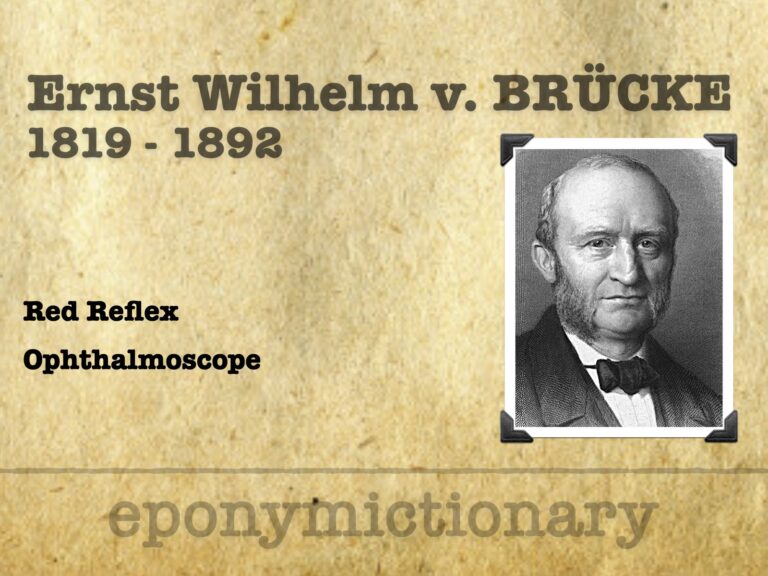
Ernst von Brücke (1819–1892) described the eye’s red reflex, paving the way for Helmholtz’s ophthalmoscope and modern retinal examination
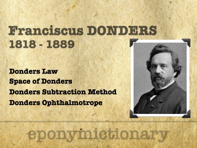
Franciscus Donders (1818–1889), Dutch ophthalmologist and physiologist, pioneered refraction studies, eye movement laws, and mental chronometry
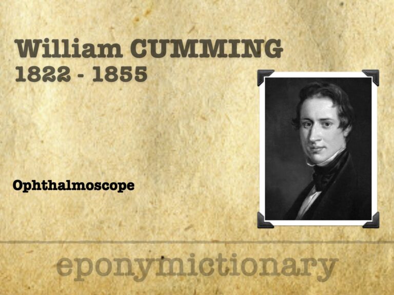
William Cumming (1822–1855), Moorfields surgeon who first observed the living eye’s luminous reflex, paving the way for Helmholtz’s ophthalmoscope.
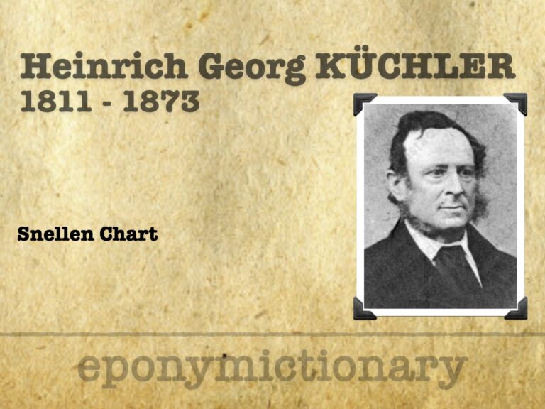
Heinrich Küchler (1811–1873): German ophthalmologist who pioneered early eye charts, advanced corneal surgery, and reformed medical and military health services
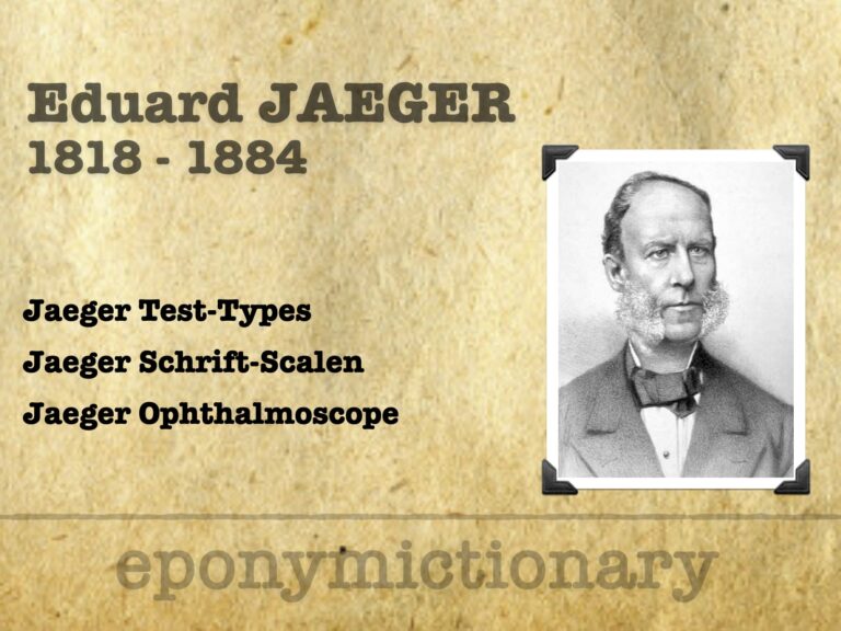
Eduard Jaeger (1818–1884), Austrian ophthalmologist; introduced Jaeger Test-Types, advanced ophthalmoscopy, and first described diabetic retinopathy
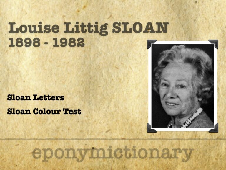
Louise L. Sloan (1898–1982) developed Sloan optotypes (LogMAR), pioneering colour vision screening, perimetry, and low-vision rehabilitation

Herman Snellen (1834–1908): Dutch ophthalmologist who created the Snellen chart and standardized visual acuity testing, transforming eye care worldwide

Today we cover lateral canthotomy and cantholysis, with a guide made in partnership with a recent publication in Australasian Emergency Care

Today we cover lateral canthotomy and cantholysis, with a guide made in partnership with a recent publication in Australasian Emergency Care
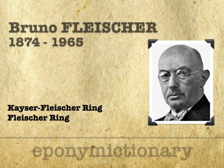
Bruno Fleischer (1874-1965) German ophthalmologist; Kayser–Fleischer ring (Wilson’s disease) and Fleischer ring in keratoconus
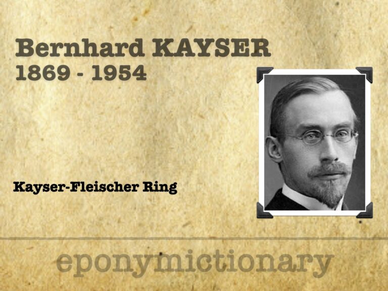
Bernhard Kayser (1869–1954) German ophthalmologist. First described the greenish-brown corneal ring now known as the Kayser–Fleischer ring in Wilson’s disease.