


ECG criteria for left ventricular hypertrophy
ECG criteria for left ventricular hypertrophy (LVH) identify an abnormal electrical phenotype associated with increased left ventricular mass but do not measure ventricular mass directly. Most criteria have high specificity but limited sensitivity with a positive result supporting LVH, while a negative ECG does not exclude anatomical hypertrophy.
Voltage should be interpreted alongside repolarisation abnormalities, atrial findings, QRS duration and axis, as well as the patient’s age, sex, body habitus and clinical context. Echocardiography or cardiac MRI is required when ventricular structure and mass need to be established.
Objective: To summarise the principal ECG criteria used to identify LVH, trace their historical development, explain the context in which each was derived, and compare their performance in selected head-to-head studies.
Clinical overview: For LVH pathophysiology, associated ECG findings, causes and worked tracings, see [Left Ventricular Hypertrophy (LVH)]
Conclusion: No ECG criterion provides a definitive measurement of anatomical LVH. Simple voltage criteria are generally specific but insensitive, while lower thresholds and composite methods increase case detection at the cost of more false-positive results. Performance varies with age, sex, body habitus, disease severity, population and reference standard.
Commonly used ECG-LVH criteria
| Named criterion | Formula | Practical interpretation |
|---|---|---|
| Sokolow–Lyon 1949 | SV1+ max(RV5,V6) ≥35mm | High specificity but low sensitivity |
| Romhilt–Estes score 1968 | Age- and sex-specific thresholds; flat/inverted T wave in V5 or V6; mm | Composite of voltage, strain, atrial abnormality, axis and conduction |
| Cornell 1987 | RaVL+SV3 >28mm (men) RaVL+SV3 >20mm (women) | Sex-specific and generally more sensitive than Sokolow–Lyon |
| Cornell voltage-duration product 1992 | [RaVL+SV3+8mm in women] × QRS duration > 2440 mm·ms | Adds ventricular activation duration to voltage |
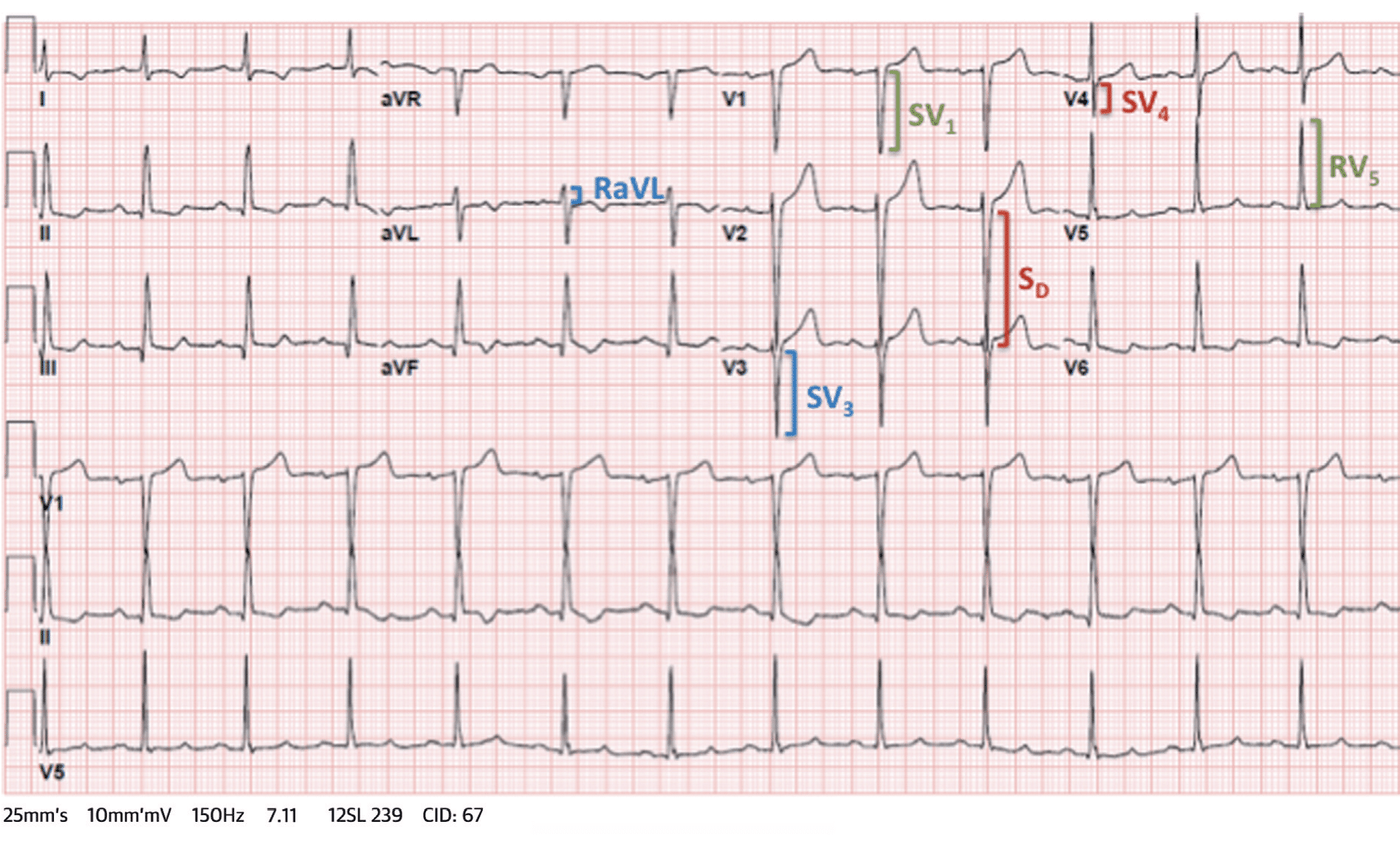
| Peguero–Lo Presti 2017 | SD+SV4 ≥ 28mm (men) SD+SV4 ≥ 23mm (women) | Uses the deepest S wave in any lead (SD) |
- Simple voltage criteria: Gubner–Ungerleider, Sokolow–Lyon, Cornell voltage, Peguero–Lo Presti
- Composite scores: Romhilt–Estes, Perugia
- Voltage–duration approaches: Cornell product
- Population-specific criteria: Araoye code
Gubner–Ungerleider criterion (1943)
Richard Gubner and Harry E. Ungerleider investigated reproducible ECG criteria for LVH in patients with hypertension and left axis deviation. They examined QRS voltage and repolarisation abnormalities to define an ECG pattern associated with advanced hypertensive heart disease. [Gubner and Ungerleider 1943]
Original definition
Gubner and Ungerleider considered LVH present when left axis deviation was accompanied by any of the following:
- Increased QRS voltage, measured as the sum of the R wave in lead I and the S wave in lead III:
- mm: LVH considered almost certain
- mm: LVH considered probable
- mm: LVH suggested
- Any perceptible ST-segment depression in lead I, including depression as small as 0.5 mm
- A low or abnormal T wave in lead I, including flattening below 1 mm, a diphasic T wave or T-wave inversion
They proposed a progression from high voltage with left axis deviation, through repolarisation “strain”, to QRS slurring, notching and widening attributed to myocardial fibrosis and evolving bundle branch block; this was a contemporary pathological interpretation rather than a validated histological classification.
Evidence and significance
The study compared 460 apparently healthy insurance applicants with left axis deviation, 380 hypertensive applicants and 100 patients with advanced hypertensive heart disease. mm occurred in approximately 1% of the presumed normal group and 52% of those with advanced disease. There was no independent anatomical LV-mass reference standard
Modern interpretation
Today, the Gubner–Ungerleider criterion generally refers only to: RI + SIII > 25 mm
The original requirement for accompanying left axis deviation and the alternative ST-segment and T-wave criteria are usually omitted from teaching. The criterion is now chiefly of historical interest and is rarely used in routine ECG interpretation.
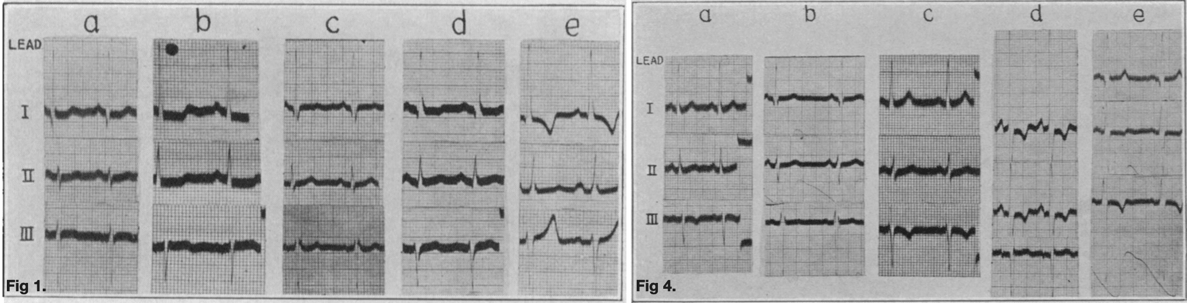
Figure 1: examples of left ventricular hypertrophy associated with left axis deviation and increased limb-lead voltage: (a) RI just exceeding normal limits; (b) deep SIII; (c) RI+SIII>25 mm; (d) markedly increased RI; and (e) markedly increased SIII.
Figure 4: examples classified as left ventricular hypertrophy and strain, combining high QRS voltage with ST-segment depression and T-wave flattening, diphasic morphology or inversion.
Sokolow–Lyon criteria (1949)
Maurice Sokolow and Thomas Lyon tried to improve recognition of early and atypical LVH using the recently introduced unipolar limb and precordial leads. They aimed to define abnormalities seen on the ECG, examine the importance of cardiac position and determine whether delayed ventricular activation provided additional diagnostic information [Sokolow and Lyon, 1949].
Original criterion
SV1 + max(RV5,V6) ≥ 35 mm
The original scheme was broader than this voltage sum and also incorporated lateral precordial voltage, ST–T abnormalities, delayed ventricular activation and cardiac orientation.
Evidence and significance
They selected 200 patients with an abnormal ECG and a clinical condition associated with increased left ventricular load, including hypertension, aortic valve disease and coarctation. After exclusions a group of 147 patients, 90% of whom had severe hypertension, were compared with 151 apparently healthy controls.
The combined SV1+ max(RV5,V6) voltage reached 35 mm in 48 of 147 patients with presumed LVH but in none of the controls. However, this was not an anatomical validation study and LVH was inferred from the clinical condition, ECG and cardiac size rather than measured by echocardiography, cardiac magnetic resonance or autopsy LV mass.
The precordial leads helped identify patients whose standard limb leads appeared normal or “atypical”. The paper reinforced that repolarisation abnormalities and delayed activation could be diagnostically important even when voltage criteria were absent.
Modern interpretation
Today, the Sokolow–Lyon criterion usually refers only to SV1 + max(RV5,V6) ≥ 35 mm
Additional findings from the same paper that remain in use are:
- RV5 or RV6 >26 mm
- RaVL >11 mm
- R-wave peak time >50 ms in V5 or V6
Sokolow–Lyon voltage remains supportive when positive but is too insensitive to exclude anatomical LVH when negative.
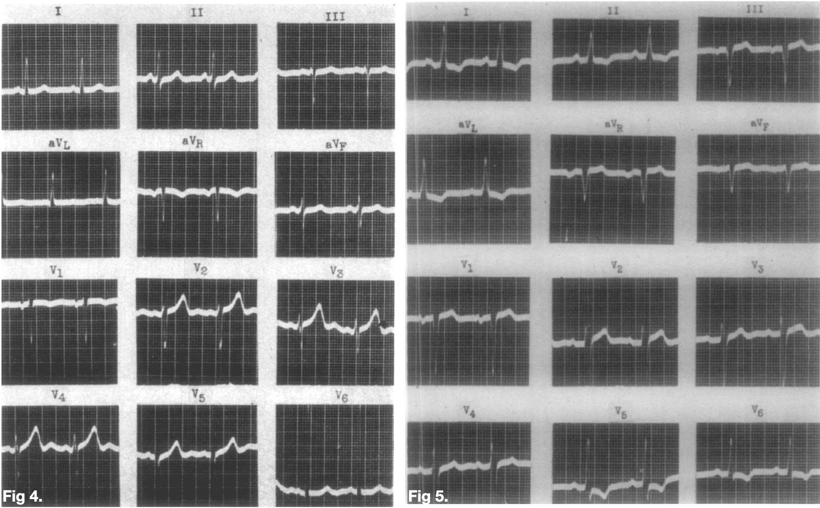
Figure 4: early LVH in a semihorizontal heart. The standard limb leads were largely unremarkable, but measured 44 mm.
Figure 5: more developed LVH with lateral ST-segment depression and asymmetric T-wave inversion—the classical secondary repolarisation or “strain” pattern.
Romhilt–Estes point score (1968)
Donald W. Romhilt and E. Harvey Estes Jr looked to improve ECG detection of LVH without sacrificing the high specificity of earlier voltage criteria. Isolated high voltage was frequently absent in anatomical LVH and could also occur in healthy young or slender patients. Their solution was to combine abnormalities from several ECG domains into a weighted point score, so that no single finding could establish the diagnosis alone.
The score was developed from earlier ECG–anatomical work, particularly Carter and Estes’ 1964 computer analysis, and incorporated the recently described terminal P-wave abnormality in V1 [Carter and Estes 1964; Romhilt and Estes, 1968].
Original criterion
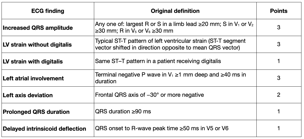
A three-point voltage or strain finding required at least one additional abnormality before the ECG could be classified as probable or definite LVH.
- ≥5 points: definite ECG LVH
- 4 points: probable ECG LVH
- Maximum score: 13 points
Evidence and significance
The score was evaluated in 150 autopsy hearts, of which 90 were classified as hypertrophied using Zeek’s body-length-adjusted heart-weight criteria. Definite LVH was identified in 57.8%, and probable or definite LVH in 62.2%. Two of 60 non-hypertrophied hearts were false positive. Romhilt–Estes shifted ECG assessment from isolated voltage towards a composite electrical phenotype incorporating voltage, repolarisation, atrial abnormality, axis and conduction.
Modern interpretation
The Romhilt–Estes score remains essentially unchanged. Modern terminology usually describes “left atrial involvement” as abnormal P-terminal force in V1 and “intrinsicoid deflection” as R-wave peak time. The digitalis distinction is now less clinically prominent.
The score remains a high-specificity, low-sensitivity composite criterion. A high score strongly supports an abnormal LV electrical phenotype, particularly when strain, conduction delay or atrial abnormality accompany voltage. A low score does not exclude anatomical LVH.
Cornell voltage criteria (1985, 1987)
Paul N. Casale, Richard B. Devereux and colleagues examined ECG variables independently associated with anatomical LV mass, develop criteria in one population and then test them prospectively in separate cohorts [Casale et al., 1985; Casale et al., 1987].
Evidence and significance
1985 – ECG variables were compared with echocardiographic LV mass in a learning cohort of 414 subjects and prospectively tested in another 129. , , T-wave amplitude in V1, age and sex provided the strongest independent information. The multivariable algorithm achieved 49% sensitivity and 93% specificity.
1987 – the simpler sex-specific voltage criterion was tested against indexed LV muscle mass in 135 autopsy patients and achieved 42% sensitivity and 96% specificity. The work established sex-specific, statistically selected lead thresholds rather than relying solely on conventional “left ventricular” leads.
Modern interpretation
Cornell voltage is named for the New York Hospital–Cornell Medical Center where the work was undertaken. The original research programme has been reduced in clinical practice to “two leads, one addition and two sex-specific thresholds”
- RaVL+ SV3 > 28 mm in men
- RaVL+ SV3 > 20 mm in women
It remains one of the most widely used simple criteria and is generally more sensitive than Sokolow–Lyon while retaining high specificity.
Framingham population study (1990)
Levy and colleagues examined how ECG criteria for LVH performed in a large community population rather than in a selected hospital, hypertensive or autopsy cohort. Their aim was to identify the patient and disease factors that influenced diagnostic sensitivity and specificity [Levy et al., 1990].
Population findings
The study included 4,684 Framingham Heart Study participants with interpretable ECGs and echocardiographic measurement of LV mass. Echocardiographic LVH was present in 290 men and 465 women.
The Framingham ECG criteria demonstrated an overall sensitivity of 6.9% and specificity of 98.8%. Sensitivity was 9.0% in men and 5.6% in women. Detection increased with age and the severity of hypertrophy, but decreased with obesity and cigarette smoking, both of which may attenuate surface QRS voltage.
Modern interpretation
The study showed that performance reported in selected hospital and autopsy cohorts did not translate directly to community screening. Sensitivity and specificity vary with the population, disease spectrum, body habitus and anatomical reference standard.
Cornell voltage–duration product (1992)
Thomas Molloy, Peter Okin, Richard Devereux and Paul Kligfield sought to improve the limited sensitivity of simple ECG voltage criteria without resorting to complex multivariable equations.
Their premise was that anatomical LVH may increase both QRS amplitude and ventricular activation duration. Neither change may cross a diagnostic threshold alone, but their combined effect could be captured by multiplying voltage by QRS duration as a practical approximation of the QRS time–voltage area [Molloy et al., 1992]
Original criterion
[RaVL+SV3+8mm in women] × QRS duration > 2440 mm·ms
Evidence and significance
The product was tested against indexed LV muscle mass in 220 autopsy patients, 95 with anatomical LVH. At matched 95% specificity, sensitivity increased from 36% for Cornell voltage to 51% for the voltage–duration product, compared with 28% for QRS duration alone and 27% for Romhilt–Estes. It extended ECG-LVH assessment from amplitude alone towards an approximation of QRS time–voltage area.
Modern interpretation
The product is generally more sensitive than Cornell voltage alone while retaining high specificity. QRS duration, however, may reflect bundle branch block, myocardial fibrosis or other conduction disease as well as increased LV mass. A negative Cornell product therefore does not exclude anatomical LVH.
Perugia score (1994, 1998)
Giuseppe Schillaci, Paolo Verdecchia and colleagues looked to improve the poor sensitivity of conventional ECG criteria in patients with essential hypertension while retaining specificity.
They combined a lower male Cornell threshold with typical strain and a definite Romhilt–Estes score. The method was developed against echocardiographic LV mass in 1994 and subsequently evaluated as a predictor of cardiovascular events in the PIUMA registry [Schillaci et al., 1994; Verdecchia et al., 1998]
Original criterion
- SV3+RaVL>24 mm in men (>20 mm in women)
or:
- a typical left ventricular strain pattern;
- a Romhilt–Estes score of at least five points.
Unlike Romhilt–Estes, the Perugia method does not assign or add points; “score” is therefore something of a misnomer. It is an OR-rule composite:
modified Cornell voltage or strain or Romhilt–Estes ≥5.
Evidence and significance
1994 – the method was developed against echocardiographic LV mass in 923 untreated White patients with essential hypertension. It achieved 34% sensitivity, 93% specificity and 73% accuracy, increasing detection principally by lowering the male Cornell threshold.
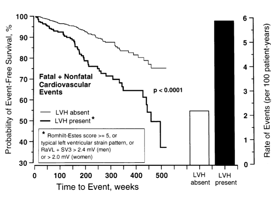
1998 – 1,717 hypertensive adults from the PIUMA registry were followed for a mean of 3.3 years. Perugia-positive LVH was present in 17.8% and was associated with event rates of 5.83 versus 2.19 per 100 patient-years. Its principal contribution was therefore the identification of a relatively prevalent hypertensive subgroup at increased cardiovascular risk.
Modern interpretation
The Perugia score is not widely used in routine reporting. It remains historically important as an attempt to combine anatomical detection with prognostic risk stratification, but its components overlap and its findings have limited transportability beyond the selected hypertensive population.
Araoye code (1996)
M. A. Araoye developed an ECG coding system for LVH in response to evidence that voltage criteria derived predominantly in European and North American populations did not perform consistently in Nigerian patients.
Rather than applying one voltage threshold to every adult, the Araoye method incorporated age and sex-specific precordial voltage limits, together with lateral repolarisation abnormality and limb-lead voltage. It was intended both to identify LVH and to express increasing ECG abnormality with a score from zero to three. [Araoye, 1996; Dada et al., 2006].
Original criterion
The code combined age- and sex-specific voltage, a flat or inverted T wave in V5 or V6, and mm. Any one component produced a positive ECG, while the number of positive components generated a code from zero to three.
Evidence and significance
In a 2006 validation involving 100 Nigerian hypertensive adults and 60 controls, Araoye achieved 71.4% sensitivity and 74.4% specificity. It was more sensitive but less specific than Cornell and Romhilt–Estes and did not significantly outperform Sokolow–Lyon as a binary test. Its importance lies in challenging the assumption that thresholds developed in one population are universally transportable.
Modern interpretation
The Araoye criterion may be summarised as positive when any of the following is present:
- SV2 + RV6 > 50 mm in men aged 15–29
- SV2 + RV6 > 40 mm in men aged ≥30
- SV2 + RV6 > 35 mm in women
or:
- flat or inverted T wave in V5 or V6
- RI > 12 mm
The Araoye code is not widely used outside the setting in which it was developed. It illustrates the need to examine the transportability of ECG criteria across populations, rather than assuming universal performance from fixed voltage thresholds.
Peguero–Lo Presti criterion (2017)
Julio Peguero, Saberio Lo Presti and colleagues tried to improve the low sensitivity of fixed-lead ECG voltage criteria for LVH. They argued that the lead showing the greatest voltage change may vary between individuals because surface voltage is affected by cardiac orientation, electrode position, body habitus, conduction and myocardial properties. Rather than measuring a predetermined R wave, the investigators examined the deepest S wave anywhere on the 12-lead ECG and combined it with the S wave in V4 [Peguero et al., 2017].
Original criterion
SD+SV4≥28 mm in men (≥23 mm in women)
with counted twice when V4 contains the deepest S wave.
Evidence and significance
The criterion was derived in 94 patients and tested in 122 additional echocardiography referrals. Sensitivity decreased from 70% in the test cohort to 57% in validation, with specificity of 89–90%. Across all 216 patients, sensitivity and specificity were 62% and 90%.
The key innovation was to identify the deepest S wave on the individual ECG rather than assume that the most informative voltage would occur in a predetermined lead. The proposed physiological explanation was that later left ventricular free-wall activation is better represented by the terminal S wave.
Modern interpretation
Peguero–Lo Presti was more sensitive than Cornell and Sokolow–Lyon in the original study, but its advantage has not been reproduced consistently in external populations. A positive result supports an abnormal LV electrical phenotype, however, a negative result does not exclude increased LV mass.
Historical and named ECG-LVH criteria
| Named criterion | Formula | Context and limitations |
|---|---|---|
| Gubner–Ungerleider 1943 | RI+SIII >25mm | Early limb-lead voltage criterion. No independent anatomical LV-mass standard; selected population with left axis deviation |
| Sokolow–Lyon 1949 | SV1+ max(RV5,V6) ≥35mm | Selected severe hypertensive population; no independent anatomical LV-mass standard; poor sensitivity in later validation |
| Romhilt–Estes score 1968 | Age- and sex-specific thresholds; flat/inverted T wave in V5 or V6; mm | Composite score incorporating voltage, strain, left atrial abnormality, axis, QRS duration and delayed intrinsicoid deflection. |
| Cornell 1987 | RaVL+SV3 >28mm (men) RaVL+SV3 >20mm (women) | Sex-specific voltage criterion developed against echocardiographic LV mass and then validated against LV mass at autopsy. |
| Cornell voltage-duration product 1992 | [RaVL+SV3+8mm in women] × QRS duration > 2440 mm·ms | Selected high-risk autopsy cohort; limited population validation; QRS duration may reflect conduction disease as well as LV mass |
| Araoye code 1996 | SV2 + RV6 flat/inverted T wave V5 or V6 RI > 12 mm Population-specific, with age- and sex-specific thresholds | Developed specifically from Nigerian ECG data to address the poor transportability of conventional thresholds to Nigerian populations. |
| Perugia score 1994, 1998 | Any of: SV3+RaVL>24mm in men or >20 mm in women; typical LV strain; Romhilt–Estes ≥5 | Components overlap; includes prognostic repolarisation and conduction abnormalities rather than measuring LV mass alone; limited population and clinical transportability |
| Peguero–Lo Presti 2017 | SD+SV4 ≥ 28mm (men) SD+SV4 ≥ 23mm (women) Deepest S wave in any lead = SD | If the deepest S wave is in V4, (S_{V4}) is counted twice. |
How do the criteria compare?
There is no universally superior ECG criterion for LVH. Comparisons are meaningful only when the criteria are applied to the same patients using the same anatomical reference standard. Even then, the apparent “winner” depends on whether priority is given to sensitivity, specificity or overall clinical utility.
Head-to-head comparisons
Cornell autopsy comparison
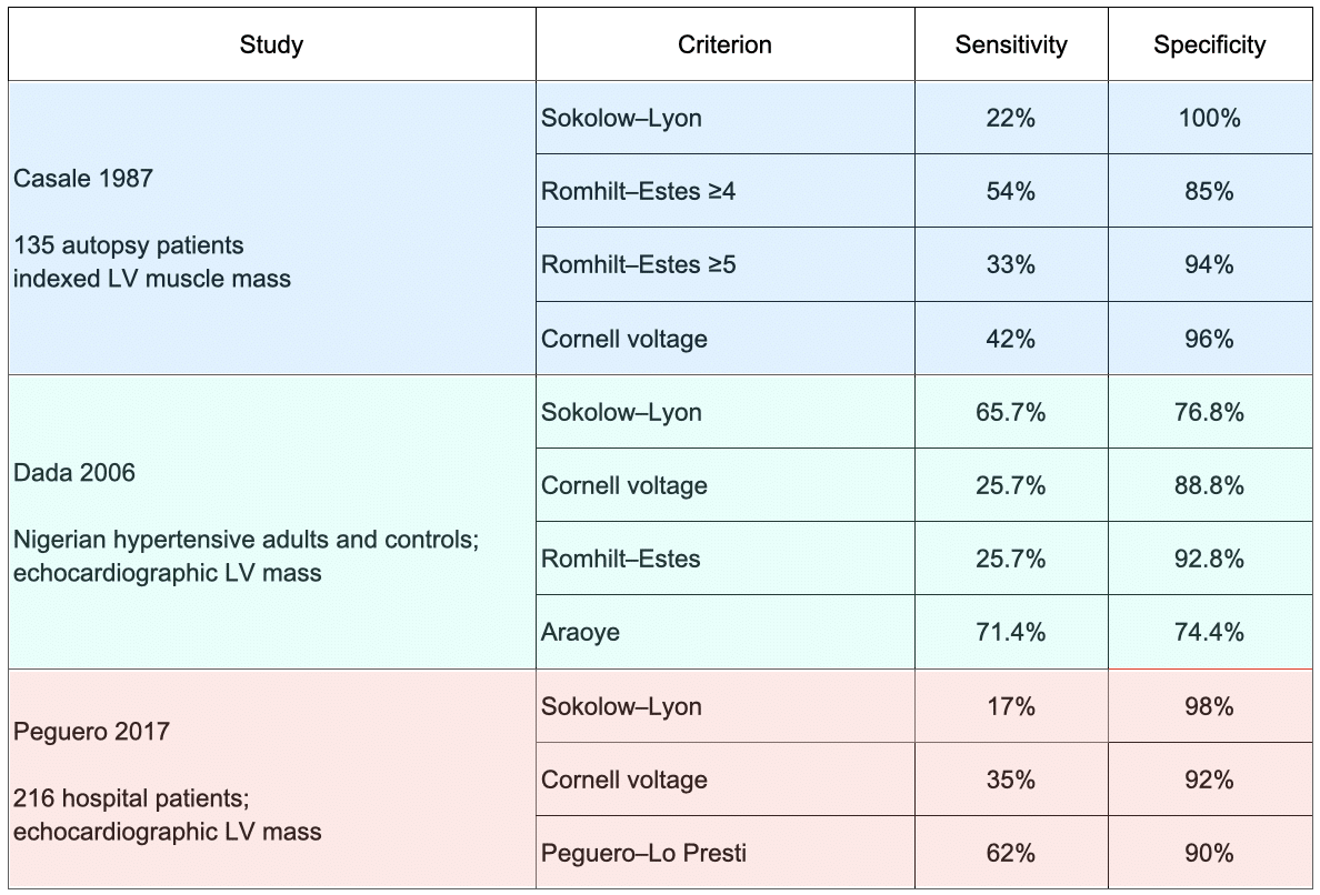
In the 1987 autopsy study, Sokolow–Lyon was completely specific but detected only 22% of anatomical LVH. Cornell voltage approximately doubled sensitivity to 42% while preserving 96% specificity. Lowering the Romhilt–Estes threshold from five to four points increased sensitivity from 33% to 54%, but reduced specificity from 94% to 85%.
Nigerian comparison
In Nigerian hypertensive adults, the balance was different. Araoye was the most sensitive criterion at 71.4%, closely followed by Sokolow–Lyon at 65.7%, but both had specificity below 80%. Cornell voltage and Romhilt–Estes were considerably less sensitive but more specific. Araoye did not significantly outperform Sokolow–Lyon as a binary diagnostic test, although increasing numbers of positive Araoye components were associated with higher blood pressure, greater wall thickness and increasing LV mass.
Peguero–Lo Presti comparison
In the combined 2017 cohorts, Peguero–Lo Presti increased sensitivity to 62%, compared with 35% for Cornell voltage and 17% for Sokolow–Lyon. Specificity remained 90%, although this was lower than Cornell at 92% and Sokolow–Lyon at 98%. These are valid internal head-to-head results, but the new thresholds were derived and tested within cohorts from the same centre; subsequent external studies have not consistently reproduced an advantage of the same magnitude
The studies therefore do not establish a single winning criterion. Instead, they illustrate a recurring trade-off:
- stringent voltage thresholds tend to preserve specificity but miss anatomical LVH;
- lower thresholds and composite criteria identify more cases but generate more false-positive results;
- demographic factors, body habitus, disease severity and population ancestry materially alter performance.
Conclusion
No ECG criterion provides a definitive measurement of anatomical LVH. Simple voltage criteria are generally specific but insensitive, while lower thresholds and composite methods increase case detection at the cost of more false-positive results. Performance varies with age, sex, body habitus, disease severity, population and reference standard.
A positive ECG criterion supports an abnormal LV electrical phenotype and may carry prognostic significance, particularly when accompanied by secondary repolarisation abnormalities. A negative ECG does not exclude increased LV mass. Echocardiography or cardiac magnetic resonance should be used when confirmation of ventricular structure or mass will influence management.
Related Topics
- Left Ventricular Hypertrophy (LVH)
- Right ventricular hypertrophy
- Left atrial enlargement
- Left bundle branch block
- Hypertrophic Cardiomyopathy (HCM)
References
Primary studies
- Gubner R, Ungerleider HE. Electrocardiographic criteria of left ventricular hypertrophy: factors determining the evolution of the electrocardiographic patterns in hypertrophy and bundle branch block. Arch Intern Med. 1943;72:196–209. [Gubner-Ungerleider]
- Sokolow M, Lyon TP. The ventricular complex in left ventricular hypertrophy as obtained by unipolar precordial and limb leads. Am Heart J. 1949 Feb;37(2):161-86 [Sokolow-Lyon criteria]
- Carter WA, Estes EH Jr. Electrocardiographic manifestations of ventricular hypertrophy; a computer study of ECG-anatomic correlations in 319 cases. Am Heart J. 1964 Aug;68:173-82.
- Romhilt DW, Estes EH Jr. A point-score system for the ECG diagnosis of left ventricular hypertrophy. Am Heart J. 1968 Jun;75(6):752-8.
- Casale PN, Devereux RB, Kligfield P, Eisenberg RR, Miller DH, Chaudhary BS, Phillips MC. Electrocardiographic detection of left ventricular hypertrophy: development and prospective validation of improved criteria. J Am Coll Cardiol. 1985 Sep;6(3):572-80.
- Casale PN, Devereux RB, Alonso DR, Campo E, Kligfield P. Improved sex-specific criteria of left ventricular hypertrophy for clinical and computer interpretation of electrocardiograms: validation with autopsy findings. Circulation. 1987 Mar;75(3):565-72. [Cornell criteria]
- Levy D, Labib SB, Anderson KM, Christiansen JC, Kannel WB, Castelli WP. Determinants of sensitivity and specificity of electrocardiographic criteria for left ventricular hypertrophy. Circulation. 1990 Mar;81(3):815-20.
- Molloy TJ, Okin PM, Devereux RB, Kligfield P. Electrocardiographic detection of left ventricular hypertrophy by the simple QRS voltage-duration product. J Am Coll Cardiol. 1992 Nov 1;20(5):1180-6. [Cornell Voltage-Duration Product]
- Schillaci G, Verdecchia P, Borgioni C, Ciucci A, Guerrieri M, Zampi I, Battistelli M, Bartoccini C, Porcellati C. Improved electrocardiographic diagnosis of left ventricular hypertrophy. Am J Cardiol. 1994 Oct 1;74(7):714-9.
- Araoye MA. Left ventricular hypertrophy by electrocardiography: A code system applicable to Negroes. Nigerian Postgraduate Medical Journal 1996; 3: 92-97.
- Verdecchia P, Schillaci G, Borgioni C, Ciucci A, Gattobigio R, Zampi I, Porcellati C. Prognostic value of a new electrocardiographic method for diagnosis of left ventricular hypertrophy in essential hypertension. J Am Coll Cardiol. 1998 Feb;31(2):383-90.
- Dada A, Adebiyi AA, Aje A, Oladapo OO, Falase AO. Comparison of Araoye’s criteria with standard electrocardiographic criteria for diagnosis of left ventricular hypertrophy in Nigerian hypertensives. West Afr J Med. 2006 Jul-Sep;25(3):179-85.
- Peguero JG, Lo Presti S, Perez J, Issa O, Brenes JC, Tolentino A. Electrocardiographic Criteria for the Diagnosis of Left Ventricular Hypertrophy. J Am Coll Cardiol. 2017 Apr 4;69(13):1694-1703.
Reviews and guidelines
- Edhouse J, Thakur RK, Khalil JM. ABC of clinical electrocardiography. Conditions affecting the left side of the heart. BMJ. 2002 May 25;324(7348):1264-7
Advanced Reading
Online
- Wiesbauer F, Kühn P. ECG Mastery: Yellow Belt online course. Understand ECG basics. Medmastery
- Wiesbauer F, Kühn P. ECG Mastery: Blue Belt online course: Become an ECG expert. Medmastery
- Kühn P, Houghton A. ECG Mastery: Black Belt Workshop. Advanced ECG interpretation. Medmastery
- Rawshani A. Clinical ECG Interpretation ECG Waves
- Smith SW. Dr Smith’s ECG blog.
- Wiesbauer F. Little Black Book of ECG Secrets. Medmastery PDF
Textbooks
- Zimmerman FH. ECG Core Curriculum. 2023
- Mattu A, Berberian J, Brady WJ. Emergency ECGs: Case-Based Review and Interpretations, 2022
- Straus DG, Schocken DD. Marriott’s Practical Electrocardiography 13e, 2021
- Brady WJ, Lipinski MJ et al. Electrocardiogram in Clinical Medicine. 1e, 2020
- Mattu A, Tabas JA, Brady WJ. Electrocardiography in Emergency, Acute, and Critical Care. 2e, 2019
- Hampton J, Adlam D. The ECG Made Practical 7e, 2019
- Kühn P, Lang C, Wiesbauer F. ECG Mastery: The Simplest Way to Learn the ECG. 2015
- Grauer K. ECG Pocket Brain (Expanded) 6e, 2014
- Surawicz B, Knilans T. Chou’s Electrocardiography in Clinical Practice: Adult and Pediatric 6e, 2008
- Chan TC. ECG in Emergency Medicine and Acute Care 1e, 2004
LITFL Further Reading
- ECG Library Basics – Waves, Intervals, Segments and Clinical Interpretation
- ECG A to Z by diagnosis – ECG interpretation in clinical context
- ECG Exigency and Cardiovascular Curveball – ECG Clinical Cases
- 100 ECG Quiz – Self-assessment tool for examination practice
- ECG Reference SITES and BOOKS – the best of the rest
ECG LIBRARY

BA MA (Oxon) MBChB (Edin) FACEM FFSEM. Emergency physician, Sir Charles Gairdner Hospital. Passion for rugby, medical history, medical education, and asynchronous learning #FOAMed evangelist. Editor-in-Chief and Webmaster of Life in the Fast lane, LITFL | On Call 4e| Eponyms | Books | Horology |

MBBS FACEM DDU (Emergency) CCPU. Emergency Physician in Melbourne, Australia. Co-Ultrasound Lead for Emergency Medicine at The Alfred Hospital. Special interests in diagnostic and procedural ultrasound, medical education, and ECG interpretation | ECG Library |