![]()
Sinus Arrhythmia
Sinus Arrhythmia Overview
Sinus rhythm with a beat-to-beat variation in the P-P interval (the time between successive P waves), producing an irregular ventricular rate.
Characteristics
- Variation in the P-P interval of more than 120 ms (3 small boxes).
- The P-P interval gradually lengthens and shortens in a cyclical fashion, usually corresponding to the phases of the respiratory cycle.
- Normal sinus P waves with a constant morphology (i.e. no evidence of premature atrial contractions).
- Constant P-R interval (i.e. no evidence of Mobitz I AV block).
Mechanism
Sinus arrhythmia is a normal physiological phenomenon, most commnonly seen in young, healthy people.
- The heart rate varies due to reflex changes in vagal tone during the different stages of the respiratory cycle.
- Inspiration increases the heart rate by decreasing vagal tone.
- With the onset of expiration, vagal tone is restored, leading to a subsequent decrease in heart rate.
- The incidence of sinus arrhythmia decreases with age, presumably due to age-related decreases in carotid distensibility and baroreceptor reflex sensitivity.
NB. “Non-respiratory” sinus arrhythmia (not linked to the respiratory cycle) is less common, typically occurs in elderly patients and is more likely to be pathological (e.g. due to heart disease or digoxin toxicity).
Differential Diagnosis
There are several other entities that cause sinus rhythm with an irregular ventricular rate:
- Frequent premature atrial contractions
- Second-degree AV block, Mobitz I (Wenckebach phenomenon)
- Type I Sinoatrial Exit Block
ECG Examples
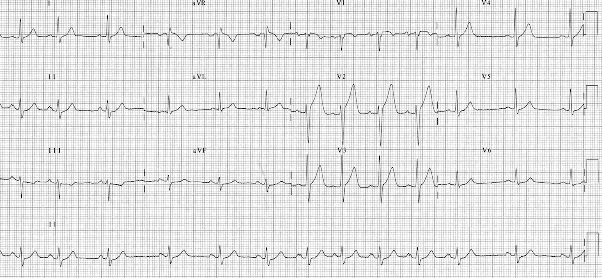
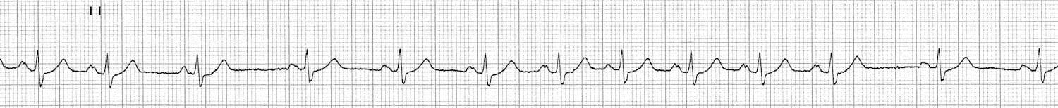
Sinus arrhythmia:
- Normal sinus P waves (upright in leads I and II) with a constant morphology — albeit with an appearance suggestive of left atrial enlargement.
- P-R interval is constant (no evidence of AV block).
- The P-P interval varies widely from 1.04 seconds (heart rate ~57 bpm) down to 0.60 seconds (heart rate ~100 bpm); a variability of over 400ms.
For irregular rhythms such as this, the ventricular rate is best estimated by multiplying the total number of complexes in the rhythm strip by 6. This gives an overall rate of 12 x 6 = 72 bpm.
Related Topics
Advanced Reading
Online
- Wiesbauer F, Kühn P. ECG Mastery: Yellow Belt online course. Understand ECG basics. Medmastery
- Wiesbauer F, Kühn P. ECG Mastery: Blue Belt online course: Become an ECG expert. Medmastery
- Kühn P, Houghton A. ECG Mastery: Black Belt Workshop. Advanced ECG interpretation. Medmastery
- Rawshani A. Clinical ECG Interpretation ECG Waves
- Smith SW. Dr Smith’s ECG blog.
- Wiesbauer F. Little Black Book of ECG Secrets. Medmastery PDF
Textbooks
- Zimmerman FH. ECG Core Curriculum. 2023
- Mattu A, Berberian J, Brady WJ. Emergency ECGs: Case-Based Review and Interpretations, 2022
- Straus DG, Schocken DD. Marriott’s Practical Electrocardiography 13e, 2021
- Brady WJ, Lipinski MJ et al. Electrocardiogram in Clinical Medicine. 1e, 2020
- Mattu A, Tabas JA, Brady WJ. Electrocardiography in Emergency, Acute, and Critical Care. 2e, 2019
- Hampton J, Adlam D. The ECG Made Practical 7e, 2019
- Kühn P, Lang C, Wiesbauer F. ECG Mastery: The Simplest Way to Learn the ECG. 2015
- Grauer K. ECG Pocket Brain (Expanded) 6e, 2014
- Surawicz B, Knilans T. Chou’s Electrocardiography in Clinical Practice: Adult and Pediatric 6e, 2008
- Chan TC. ECG in Emergency Medicine and Acute Care 1e, 2004
LITFL Further Reading
- ECG Library Basics – Waves, Intervals, Segments and Clinical Interpretation
- ECG A to Z by diagnosis – ECG interpretation in clinical context
- ECG Exigency and Cardiovascular Curveball – ECG Clinical Cases
- 100 ECG Quiz – Self-assessment tool for examination practice
- ECG Reference SITES and BOOKS – the best of the rest
ECG LIBRARY
Emergency Physician in Prehospital and Retrieval Medicine in Sydney, Australia. He has a passion for ECG interpretation and medical education | ECG Library |