![]()
Left Anterior Fascicular Block (LAFB)
In left anterior fascicular block (LAFB), impulses are conducted to the left ventricle via the posterior fascicle, producing characteristic ECG changes

![]()
In left anterior fascicular block (LAFB), impulses are conducted to the left ventricle via the posterior fascicle, producing characteristic ECG changes
Left posterior fascicular block LPFB (left posterior hemiblock), impulses are conducted to the left ventricle via the left anterior fascicle
A review of the ECG features of right ventricular outflow tract tachycardia (RVOT), a type of VT, with example ECGs.
Isolated low serum Mg levels are associated with atrial depolarisation and ventricular repolarisation abnormalities, predisposing to ventricular arrhythmias
Mixed pattern of RBBB in precordial leads and LBBB in limb leads, with a higher rate of progression to complete heart block than typical bifascicular block
Trifascicular block (TFB) refers to the presence of conducting disease in all three fascicles: RBBB, LAFB, LPFB. LITFL ECG Library
ECG Axis. Hexaxial QRS Axis analysis for dummies. Quick and easy method of estimating ECG axis with worked examples and differential diagnoses

Dressler beat: Specifically a 'ventricular fusion beat' in the presence of paroxysmal ventricular tachycardia. Wide complex tachycardia with VT
Left Bundle Branch Block LBBB - normal direction of septal depolarisation is reversed (becomes right to left), as the impulse spreads
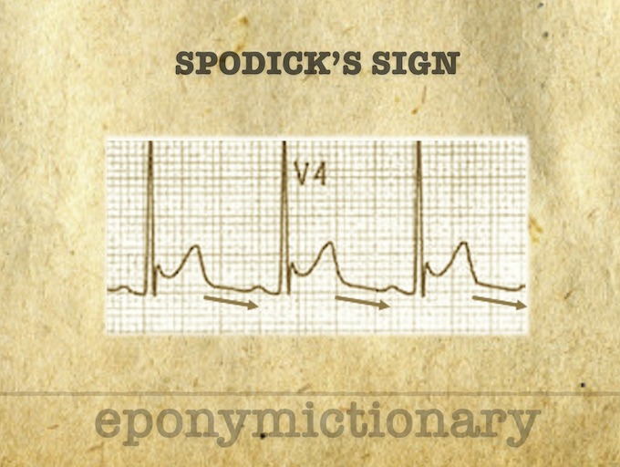
Spodick Sign: Stage I Pericarditis, a downsloping of the TP line. Described 1974 by American Cardiologist, David H Spodick (1927 – 2019)
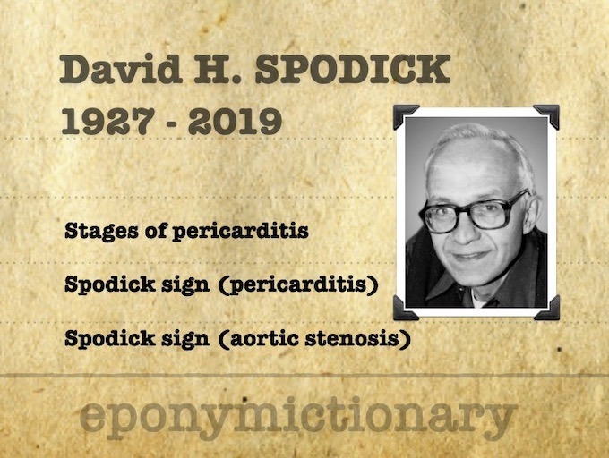
David H Spodick (1927 – 2019) was an American Cardiologist. Eponymously affiliated with Spodick's sign in acute pericarditis (1974)
The OMI/NOMI paradigm: Better recognising patients with acute ischaemia that will respond to Percutaneous Coronary Intervention (PCI)