
![]()
ECG Case 035
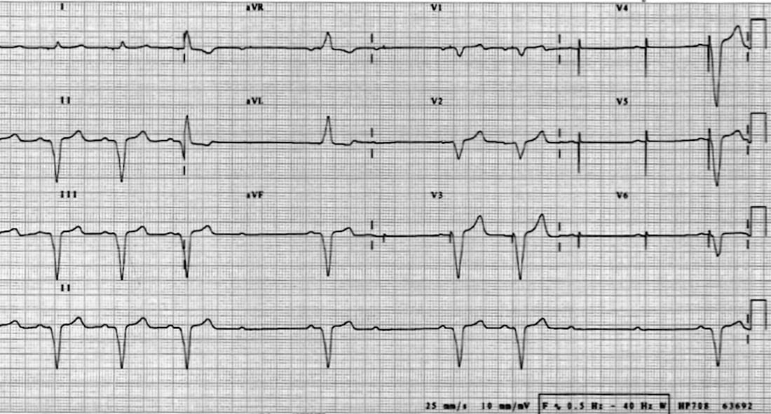
80-year old patient presenting with syncope. Describe the ECG.
Describe and interpret this ECG
ECG ANSWER and INTERPRETATION
On first glance this ECG could easily be mistaken for an example of Mobitz II AV block — there are intermittent non-conducted P waves with a constant PR interval. However, regular pacing spikes can be seen following the P waves in leads V3-6.
This is an example of pacemaker malfunction, with intermittent failure to capture:
- Regular P waves are seen at ~65 bpm
- Each P wave is following by a pacing spike (best seen in V3-6, subtle pacing spikes also present in I, aVR, V1). This indicates that atrial sensing is intact. NB. Pacing spikes will typically not be seen in all 12 leads
- Some of the pacing spikes are followed by typical ventricular-paced complexes. The LBBB morphology indicates that the pacing lead is in the right ventricle — the heart depolarises from right to left in the same way as LBBB. Also note the negative concordance in V1-6 (all QRS complexes are negative). This is often quoted as a feature of ventricular tachycardia, but simply indicates that the rhythm is arising from the anterior surface of the right ventricle — the heart is depolarising away from the V1-6 electrodes
- Several of the P waves / pacing spikes are not followed by QRS complexes, producing a ventricular rate of ~ 40 bpm
- Quite worryingly, there does not seem to be any native ventricular activity kicking in when the heart rate drops. The second half of the rhythm strip shows two sequential non-conducted P waves with no evidence of any escape rhythm. This suggests the presence of underlying complete heart block with inadequate escape mechanisms and significant risk of ventricular standstill
CLINICAL PEARLS
Failure to Capture
The problem here is failure of the pacemaker to “capture” (depolarise) the ventricular myocardium. Causes of this include:
- Pacemaker lead fracture or migration (e.g. due to Twiddler’s syndrome)
- Refractory myocardium — due to electrolyte abnormality (esp. hyperkalaemia) or myocardial ischaemia
References
Further Reading
- Wiesbauer F, Kühn P. ECG Mastery: Yellow Belt online course. Understand ECG basics. Medmastery
- Wiesbauer F, Kühn P. ECG Mastery: Blue Belt online course: Become an ECG expert. Medmastery
- Kühn P, Houghton A. ECG Mastery: Black Belt Workshop. Advanced ECG interpretation. Medmastery
- Rawshani A. Clinical ECG Interpretation ECG Waves
- Smith SW. Dr Smith’s ECG blog.
- Wiesbauer F. Little Black Book of ECG Secrets. Medmastery PDF
TOP 100 ECG Series
Emergency Physician in Prehospital and Retrieval Medicine in Sydney, Australia. He has a passion for ECG interpretation and medical education | ECG Library |

MBBS FACEM DDU (Emergency) CCPU. Emergency Physician in Melbourne, Australia. Co-Ultrasound Lead for Emergency Medicine at The Alfred Hospital. Special interests in diagnostic and procedural ultrasound, medical education, and ECG interpretation. Editor of the LITFL ECG Library.
