![]()
Ventricular Escape Rhythm
Definition
Ventricular Escape Rhythm: A ventricular rhythm with a rate of 20-40 bpm.
- QRS complexes are broad (≥ 120 ms) and may have a LBBB or RBBB morphology.
- Also known as Idioventricular escape rhythm
Mechanism
Pacemaker cells are found at various sites throughout the conducting system, with each site capable of independently sustaining the heart rhythm. The rate of spontaneous depolarisation of pacemaker cells decreases down the conducting system:
- SA node (60-100 bpm)
- Atria (< 60 bpm)
- AV node (40-60 bpm)
- Ventricles (20-40 bpm)
Under normal conditions, subsidiary pacemakers are suppressed by the more rapid impulses from above (i.e. sinus rhythm). Junctional and ventricular escape rhythms arise when the rate of supraventricular impulses arriving at the AV node or ventricle is less than the intrinsic rate of the ectopic pacemaker.
Causes
Conditions leading to the emergence of a junctional or ventricular escape rhythm include:
- Severe sinus bradycardia
- Sinus arrest
- Sino-atrial exit block
- High-grade second degree AV block
- Third degree AV block
- Hyperkalaemia
- Drugs: beta-blocker, calcium-channel blocker or digoxin poisoning
ECG Examples
Example 1

Sinus arrest with a ventricular escape rhythm
- Sinus pause / arrest (there is a single P wave visible on the 6-second rhythm strip).
- Broad complex escape rhythm with a LBBB morphology at a rate of 25 bpm.
- The LBBB morphology (dominant S wave in V1) suggests a ventricular escape rhythm arising from the right bundle branch.
Example 2

Complete heart block with a ventricular escape rhythm
- Sinus rhythm with 3rd degree AV block.
- Broad complex escape rhythm at around 27 bpm.
- The RBBB (dominant R wave in V1) + left posterior fascicular block (right axis deviation) morphology suggests a ventricular escape rhythm arising from the left anterior fascicle.
Example 3
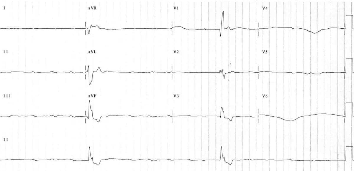
Complete heart block with a ventricular escape rhythm
- Sinus rhythm with 3rd degree AV block.
- Extremely slow broad complex escape rhythm (around 15 bpm).
- The RBBB morphology (dominant R wave in V1) indicates a ventricular escape rhythm arising somewhere within the left bundle branch.
Related Topics
Advanced Reading
Online
- Wiesbauer F, Kühn P. ECG Mastery: Yellow Belt online course. Understand ECG basics. Medmastery
- Wiesbauer F, Kühn P. ECG Mastery: Blue Belt online course: Become an ECG expert. Medmastery
- Kühn P, Houghton A. ECG Mastery: Black Belt Workshop. Advanced ECG interpretation. Medmastery
- Rawshani A. Clinical ECG Interpretation ECG Waves
- Smith SW. Dr Smith’s ECG blog.
- Wiesbauer F. Little Black Book of ECG Secrets. Medmastery PDF
Textbooks
- Zimmerman FH. ECG Core Curriculum. 2023
- Mattu A, Berberian J, Brady WJ. Emergency ECGs: Case-Based Review and Interpretations, 2022
- Straus DG, Schocken DD. Marriott’s Practical Electrocardiography 13e, 2021
- Brady WJ, Lipinski MJ et al. Electrocardiogram in Clinical Medicine. 1e, 2020
- Mattu A, Tabas JA, Brady WJ. Electrocardiography in Emergency, Acute, and Critical Care. 2e, 2019
- Hampton J, Adlam D. The ECG Made Practical 7e, 2019
- Kühn P, Lang C, Wiesbauer F. ECG Mastery: The Simplest Way to Learn the ECG. 2015
- Grauer K. ECG Pocket Brain (Expanded) 6e, 2014
- Surawicz B, Knilans T. Chou’s Electrocardiography in Clinical Practice: Adult and Pediatric 6e, 2008
- Chan TC. ECG in Emergency Medicine and Acute Care 1e, 2004
LITFL Further Reading
- ECG Library Basics – Waves, Intervals, Segments and Clinical Interpretation
- ECG A to Z by diagnosis – ECG interpretation in clinical context
- ECG Exigency and Cardiovascular Curveball – ECG Clinical Cases
- 100 ECG Quiz – Self-assessment tool for examination practice
- ECG Reference SITES and BOOKS – the best of the rest
ECG LIBRARY
Emergency Physician in Prehospital and Retrieval Medicine in Sydney, Australia. He has a passion for ECG interpretation and medical education | ECG Library |

MBBS DDU (Emergency) CCPU. Adult/Paediatric Emergency Medicine Advanced Trainee in Melbourne, Australia. Special interests in diagnostic and procedural ultrasound, medical education, and ECG interpretation. Co-creator of the LITFL ECG Library. Twitter: @rob_buttner