


Shenton Line
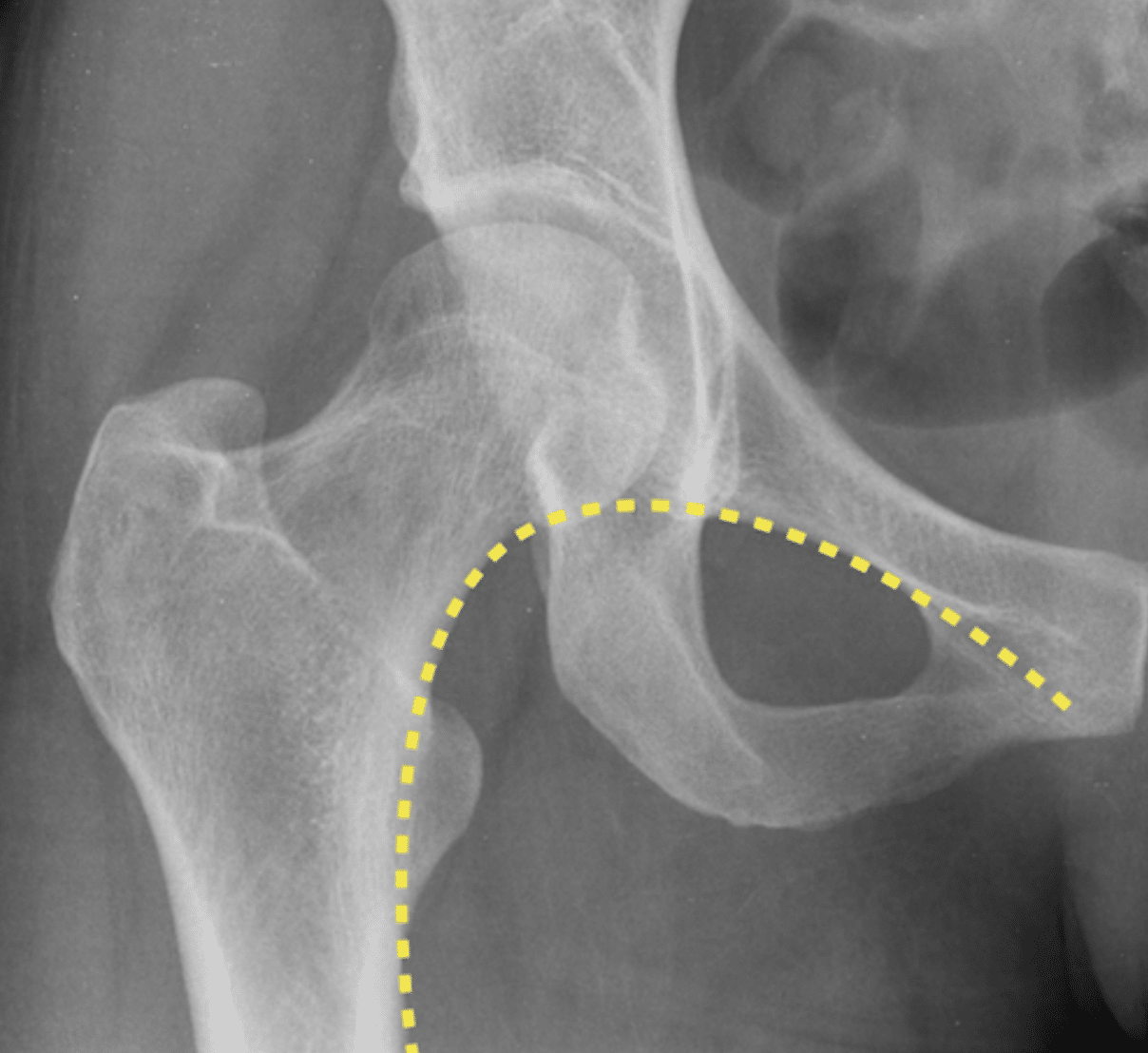
Shenton line (Shenton arc; Shenton–Ménard line; cervico-obturator arch) is a smooth imaginary radiographic curve used to assess alignment of the hip on an anteroposterior pelvis or hip radiograph. It follows the inferior border of the superior pubic ramus / superior margin of the obturator foramen and continues into the inferomedial border of the femoral neck.
In the normally aligned hip the two contours form a smooth, approximately continuous arc. Loss of continuity, a step or asymmetry between the two sides suggests displacement of the proximal femur relative to the pelvis. Abnormality may occur with developmental hip dysplasia or subluxation, hip dislocation, displaced femoral-neck fracture, pelvic injury and destructive disease of the hip.
The line is best considered a screening sign of alignment rather than a diagnosis in itself. Shenton cautioned that preservation of the curve did not exclude pathology. An undisplaced femoral-neck fracture, for example, may leave the line intact.
History
1902 – February 6: Edward Warren Hine Shenton (1872–1955) presented A system of radiography to the Roentgen Society. While discussing the particular difficulty of diagnosing fractures around the hip, he wrote of a line whose “broken continuity” pointed to trouble in the hip region and again defined the curve by the upper obturator margin and inner femoral neck.
1902 – February 14: At the second meeting of the British Electrotherapeutic Society, Shenton presented A diagnostic line about the hip joint to the British Electrotherapeutic Society. He described an imaginary arch formed by the upper margin of the obturator foramen and inner margin of the femoral neck. Loss of continuity suggested hip displacement or disease such as dislocation, femoral-neck fracture, pelvic fracture and disease of the joint surfaces. Shenton cautioned that an intact line did not exclude an undisplaced fracture.
1907 – French orthopaedic surgeon Victor Ménard (1854-1934) independently described the same cervico-obturator relationship in Étude sur la coxalgie. He observed that the curved line corresponding to the superior half of the obturator foramen appeared to continue with the concave medial border of the femoral neck, forming a very regular arch or “cintre très régulier”. Loss of arch symmetry allowed subtle superior displacement of the femoral head and neck to be recognised. The sign subsequently became known in French literature as the ligne de Ménard or cintre cervico-obturateur.
Original
English
A l’état normal, on peut remarquer que la ligne courbe, répondant à la moitié supérieure du trou ovale, paraît se continuer, en dehors de l’ischion, avec la ligne concave limitant la face interne du col en formant un cintre très régulier. Il semble qu’il s’agisse d’un seul arc interrompu par l’ischion.
Cette donnée permet d’apprécier, au premier coup d’œil, la plus légère élévation de la tête et du col du fémur du côté malade : la moitié externe du cintre s’est relevée, alors qu’une régularité parfaite est conservée du côté sain.
Un léger degré d’élévation de la tête fémorale est encore traduit, d’une manière frappante, par l’élargissement de l’espace articulaire en bas et en dedans, au niveau de l’ischion, alors que cet espace est rétréci en haut sous le sourcil (Voy. pl. IV). A un degré plus accentué, l’ascension est frappante au premier coup d’œil ; on la mesure avec le compas en prenant pour point de repère la crête iliaque du côté malade et du côté sain.
Comme il s’agit d’un déplacement suivant la longueur du sujet, en hauteur sur le dessin, l’image n’est pas sujette à erreur. On voit exactement sur ces épreuves à quel degré la tête s’élève et empiète sur la saillie du sourcil comment ces deux surfaces, dans ce rapport anormal, se sont ulcérées réciproquement. Seule la radiographie permet de faire la distinction un peu subtile entre ces deux degrés de luxation incomplète, que Lannelongue a désignés par les termes d’empiétement et de chevauchement.
Ménard Étude sur la coxalgie 1907
In the normal state, it may be observed that the curved line corresponding to the upper half of the obturator foramen appears to continue, lateral to the ischium, with the concave line bounding the medial surface of the femoral neck, forming a very regular arch. It appears to constitute a single arc interrupted by the ischium.
This relationship makes it possible, at a glance, to recognise even the slightest elevation of the femoral head and neck on the affected side: the lateral half of the arch has risen, while perfect regularity is preserved on the healthy side.
A slight degree of elevation of the femoral head is also strikingly demonstrated by widening of the joint space inferiorly and medially, at the level of the ischium, while the space is narrowed superiorly beneath the acetabular roof (see Plate IV). With greater displacement, the upward migration is obvious at first glance and can be measured with callipers, using the iliac crest on the affected and unaffected sides as reference points.
Because this displacement occurs along the long axis of the patient, vertically on the image, the radiographic appearance is not subject to error. These images show precisely the degree to which the head rises and encroaches upon the acetabular roof…
Ménard Étude sur la coxalgie 1907
Ménard illustrated the associated asymmetry in Plate IV.
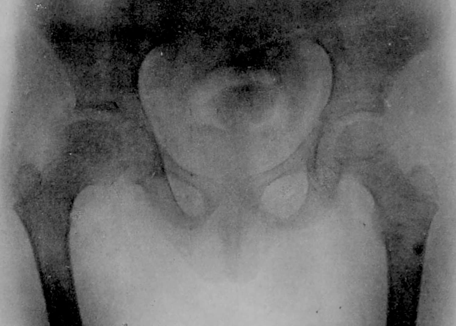
1908 – Radiologist Maxime Ménard (1872–1926) defended his thesis Le bassin normal en radiographie at the Faculté de Médecine de Paris. Later historical literature has attributed an independent description of the Shenton–Ménard line to this thesis. However, examination of the thesis did not identify a description of the cervico-obturator arch. Attribution to Maxime appears to be a biographical misattribution.
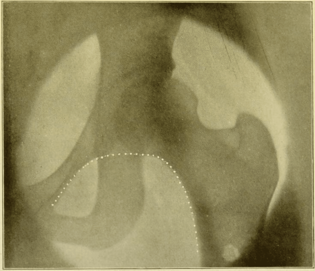
1911 – Shenton published Disease in Bone and Its detection by the X-Rays, containing the earliest illustration located of his diagnostic hip line. He emphasised its value in identifying subtle displacement, including tuberculous disease and congenital dislocation.
1920 – Norwegian surgeon Halfdan Sundt (1873-1951) first published the eponymous term “Shenton’s Line (Sh. L.)” in his thesis on Calvé–Legg–Perthes disease. The original passage has not yet been directly located, but Carl Renner quoted a translation in 1925
Original
English
Das Verhältnis des Gelenkspaltes wird am besten kontrolliert durch Shenton’s Line (Sh. L.); ein Bruch in ihr bedeutet ein Hinaufrücken des Femurendes in die Azetabularhöhle, oder ein mehr oder weniger vollständiges Herausrücken desselben.
The relationship of the joint space is best assessed using Shenton’s line (Sh. L.); a break in this line indicates an upward displacement of the femoral head into the acetabulum or a more or less complete displacement out of it.
1925 – Carl Renner published Die Shenton’sche Linie in Zentralblatt für Chirurgie. He promoted the line as a useful radiographic control of hip alignment and illustrated its disruption in tuberculous coxitis, congenital subluxation and Perthes disease. Renner noted that the line was already present in some English radiographic atlases and specifically cited Sundt’s 1920 monograph, while observing that it had received little attention in German literature.
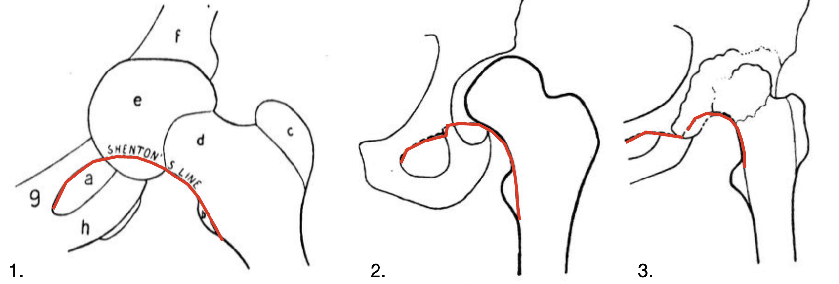
2005 – Campbell included the Shenton arc among the standard radiographic lines used to assess the pelvis and hip. He defined it as the smooth curvilinear continuation between the medial femoral neck and undersurface of the superior pubic ramus.
2011 – Rhee et al assessed the reliability of Shenton’s line in 128 skeletally mature hips and reported high observer agreement; for acetabular dysplasia the mean sensitivity was 83.3% and specificity 98.4%, supporting its value as a marker of superior femoral-head subluxation.
Associated Persons
- Edward Warren Hine Shenton (1872–1955)
- Victor Ménard (1854-1934)
- Maxime Ménard (1872–1926)
- Halfdan Sundt (1873-1951)
- Heinrich Hilgenreiner (1870-1953) and the Hilgenreiner line
- George Perkins (1892-1979) and the Perkins line
- Dietrich Tönnis (1927-2010) and the Tönnis angle
Alternative names
- cervico-obturator arc, cintre fémoropubie, cintre cervico-obturateur
- Shenton–Ménard line, Ménard-Shenton line
Eponymythology
Shenton described the cervico-obturator arc in Britain in 1902. Victor Ménard (1854-1934) independently described the same radiographic relationship in France in 1907, and the French terms ligne de Ménard and cintre cervico-obturateur reflect this parallel description.
In modern historical literature, however, the eponym is commonly attributed to French radiologist Maxime Ménard (1872–1926). This is usually referenced to his 1908 thesis Le bassin normal en radiographie.
Examination of the primary sources identifies the description in Victor Ménard’s Étude sur la coxalgie (1907 p.255), while no equivalent passage has been found in Maxime Ménard’s thesis. The later attribution to Maxime may represent a bibliographical conflation. The combination of a shared surname, overlapping radiological interests and references abbreviated to “M. Ménard” provides a plausible route by which Victor Ménard’s 1907 observation became attached to Maxime Ménard and his 1908 thesis.
References
Historical references
- Shenton EWH. A diagnostic line about the hip joint. Journal of Physical Therapeutics 1902;3:110-12.
- Shenton EWH. A system of radiography. Archives of the Roentgen Ray, 1902; 6(4): 62-70.
- Ménard V. Luxation incomplète. Empiétement. Chevauchement. In: Étude sur la coxalgie. 1907: 255
- Ménard P-F-M M. Le bassin normal en radiographie. 1908
- Shenton EWH. Disease in Bone and Its detection by the X-Rays. 1911: 41-43
- Sundt H. Undersøkelser over malum coxæ Calvé-Legg-Perthes: et bidrag til vort kjendskab til de ikke tuberkuløse hofteledsygdomme særlig i barnealderen. Kristiania: Jacob Dybwad, 1920
- Sundt H. Malum Coxae Calvé-Legg-Perthes. Zentralbl Chir. 1920; 47.1(22):538-539.
- Renner C. Die Shenton’sche Linie. Zentralblatt für Chirurgie 1925; 52: 2875-6
Eponymous term review
- Campbell SE. Radiography of the hip: lines, signs, and patterns of disease. Semin Roentgenol. 2005 Jul;40(3):290-319.
- Jones DH. Shenton’s line. J Bone Joint Surg Br. 2010 Sep;92(9):1312-5.
- Rhee PC et al. The Shenton line in the diagnosis of acetabular dysplasia in the skeletally mature patient. J Bone Joint Surg Am. 2011 May;93 Suppl 2:35-9.
- Bi AS, Carter C, Price AE, Litrenta J, Karamitopoulos M, Castañeda PG. A history of eponym usage in hip and pelvis radiography part 1: the paediatric hip. Hip Int. 2023 Mar;33(2):136-143.
- Bi AS, Ryan MK, Castañeda PG, Youm T. A history of eponym usage in hip and pelvis radiography part 2: the adult hip. Hip Int. 2023 Mar;33(2):144-151
- Vanhoenacker FM, Feydy A. History Page: Leaders in MSK Radiology Maxime Ménard, 1872-1926. Semin Musculoskelet Radiol. 2023 Aug;27(4):487-488.
- Jones J. Pelvis annotated x-ray. Radiopaedia
- Kyle E, Cadogan M. Hip and pelvic lines and arcs. LITFL
eponymictionary
the names behind the name

MBChB University of Manchester, PG Dip (SEM) University of Bath. Keen interest in Sport and Exercise Medicine. Currently working in emergency medicine in Perth, WA and intend to train as an ACSEP Registrar.

BA MA (Oxon) MBChB (Edin) FACEM FFSEM. Emergency physician, Sir Charles Gairdner Hospital. Passion for rugby, medical history, medical education, and asynchronous learning #FOAMed evangelist. Editor-in-Chief and Webmaster of Life in the Fast lane, LITFL | On Call 4e| Eponyms | Books | Horology |