
![]()
Low QRS Voltage
Diagnostic criteria
The QRS is said to be low voltage when:
- The amplitudes of all the QRS complexes in the limb leads are < 5 mm; or
- The amplitudes of all the QRS complexes in the precordial leads are < 10 mm
Mechanisms
Low voltage is produced by:
- The “damping” effect of increased layers of fluid, fat or air between the heart and the recording electrode
- Loss of viable myocardium
- Diffuse infiltration or myxoedematous involvement of the heart
Causes
The most important cause is massive pericardial effusion, which produces a triad of:
- Low voltage
- Tachycardia
- Electrical alternans
Patients with this triad need to be immediately assessed for clinical or echocardiographic evidence of tamponade.
Other causes of low voltage include:
- Fluid: Pericardial effusion; Pleural effusion
- Fat: Obesity
- Air: Emphysema; Pneumothorax
- Infiltrative / Connective Tissue Disorders
- Myxoedema
- Infiltrative myocardial diseases — i.e. restrictive cardiomyopathy due to amyloidosis, sarcoidosis, haemochromatosis
- Constrictive pericarditis
- Scleroderma
- Loss of viable myocardium:
- Previous massive MI
- End-stage dilated cardiomyopathy
ECG Examples
Example 1
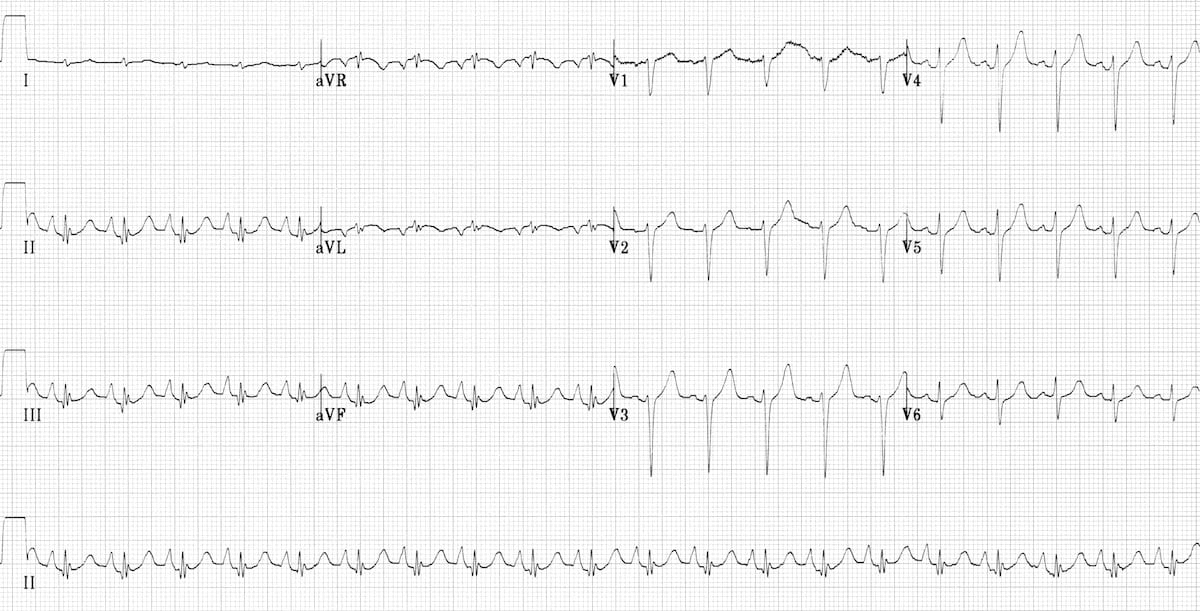
Massive Pericardial Effusion:
- Characteristc triad of tachycardia, low voltage QRS and electrical alternans
Example 2
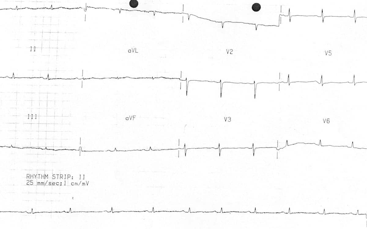
Prior Massive Anterior MI:
- Low QRS voltage in V1-6. This diffuse loss of R wave height suggests extensive myocardial loss from a prior anterior MI.
- There is also biphasic anterior T waves (Wellens syndrome) indicating new critical occlusion of the LAD artery
Example 3
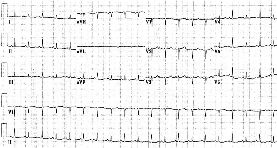
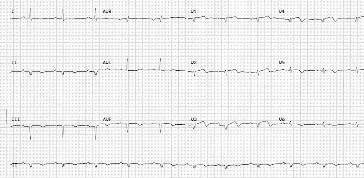
Emphysema:
- Low voltages in the limb leads is classically seen in patients with emphysema.
- Other assocaited ECG features of emphysema include:
- Right axis deivation
- Peaked P waves (P pulmonale)
- Clockwise rotation (persistent S wave in V6)
Related Topics
References
- Ultrasound Podcast – How to diagnose pericardial tamponade on bedside echo, Part 1 and Part 2 (video lessons)
- Dr Smith’s ECG Blog – Differential diagnosis of low QRS voltage (case discussion)
Advanced Reading
Online
- Wiesbauer F, Kühn P. ECG Mastery: Yellow Belt online course. Understand ECG basics. Medmastery
- Wiesbauer F, Kühn P. ECG Mastery: Blue Belt online course: Become an ECG expert. Medmastery
- Kühn P, Houghton A. ECG Mastery: Black Belt Workshop. Advanced ECG interpretation. Medmastery
- Rawshani A. Clinical ECG Interpretation ECG Waves
- Smith SW. Dr Smith’s ECG blog.
- Wiesbauer F. Little Black Book of ECG Secrets. Medmastery PDF
Textbooks
- Zimmerman FH. ECG Core Curriculum. 2023
- Mattu A, Berberian J, Brady WJ. Emergency ECGs: Case-Based Review and Interpretations, 2022
- Straus DG, Schocken DD. Marriott’s Practical Electrocardiography 13e, 2021
- Brady WJ, Lipinski MJ et al. Electrocardiogram in Clinical Medicine. 1e, 2020
- Mattu A, Tabas JA, Brady WJ. Electrocardiography in Emergency, Acute, and Critical Care. 2e, 2019
- Hampton J, Adlam D. The ECG Made Practical 7e, 2019
- Kühn P, Lang C, Wiesbauer F. ECG Mastery: The Simplest Way to Learn the ECG. 2015
- Grauer K. ECG Pocket Brain (Expanded) 6e, 2014
- Surawicz B, Knilans T. Chou’s Electrocardiography in Clinical Practice: Adult and Pediatric 6e, 2008
- Chan TC. ECG in Emergency Medicine and Acute Care 1e, 2004
LITFL Further Reading
- ECG Library Basics – Waves, Intervals, Segments and Clinical Interpretation
- ECG A to Z by diagnosis – ECG interpretation in clinical context
- ECG Exigency and Cardiovascular Curveball – ECG Clinical Cases
- 100 ECG Quiz – Self-assessment tool for examination practice
- ECG Reference SITES and BOOKS – the best of the rest
ECG LIBRARY
Emergency Physician in Prehospital and Retrieval Medicine in Sydney, Australia. He has a passion for ECG interpretation and medical education | ECG Library |

MBBS FACEM DDU (Emergency) CCPU. Emergency Physician in Melbourne, Australia. Co-Ultrasound Lead for Emergency Medicine at The Alfred Hospital. Special interests in diagnostic and procedural ultrasound, medical education, and ECG interpretation. Editor of the LITFL ECG Library.
Regarding ECG #2, can we diagnose Wellens when there is q waves and loss of R wave progression?
Very helpful💯🙏🙏🙏🙏