![]()
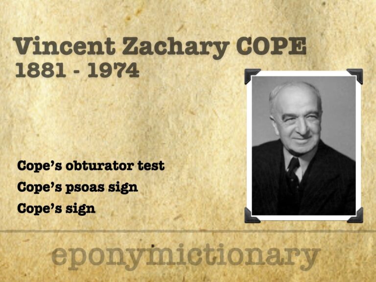
Vincent Cope
Sir Vincent Zachary Cope (1881 – 1974) was a British physician and surgeon. Eponymously linked with Cope Psoas test and obturator test.

![]()
Sir Vincent Zachary Cope (1881 – 1974) was a British physician and surgeon. Eponymously linked with Cope Psoas test and obturator test.
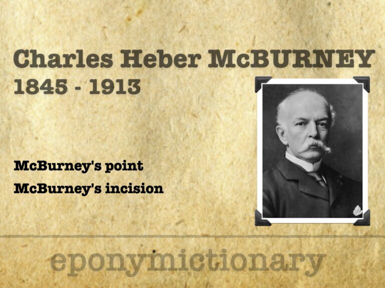
Charles Heber McBurney (1845 – 1913) was an American surgeon. Most famous for McBurney's point (1889) and McBurney's incision (1894) Medical Eponym.

James Sherren (1872-1945) British General surgeon. Eponym: Sherren's triangle - area of hyperaesthesia associated with appendicitis
Abdominal CT reporting: The basis of both image review and reporting is that of a search pattern.
Abdominal CT: The bones are often one of the last items on the reporting checklist for abdominal CT, but they still deserve our careful attention.
Abdominal CT: body wall. Evaluating the body wall - Musculature, Subcutaneous fat and skin
Abdominal CT. Evaluating for abnormal or enlarged lymph nodes is an essential part of any abdominal CT, particularly when staging cancer.
Abdominal CT. Checking the abdominal and pelvic venous systems. Review of the systemic and portal venous system
Abdominal CT. Checking the abdominal arteries. The Coeliac axis, Superior and inferior mesenteric arteries, renal arteries, and Common iliac arteries
Abdominal CT. Imaging the large bowel. in particular the location and identification of the appendix
Abdominal CT: appendicitis. Identifying acute appendicitis, perforated appendix and abscess formation
Abdominal CT: Common Terms used to describe pathology seen on CT images. Including thickened, fat stranding and fluid