Abdominal CT: peptic ulcer disease
Abdominal CT: peptic ulcer disease. Recognising peptic ulcer disease and key CT findings to increase radiological suspicion

Abdominal CT: peptic ulcer disease. Recognising peptic ulcer disease and key CT findings to increase radiological suspicion

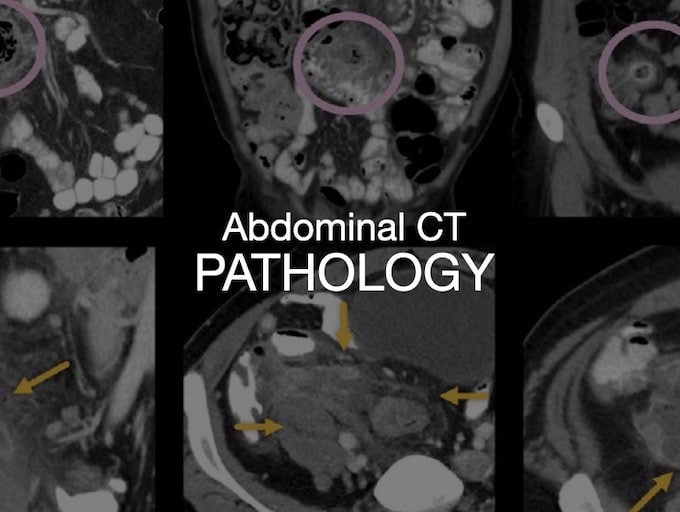
Abdominal CT: enteritis and colitis. Identifying enteritis and colitis, we review the most common presentations of gastrointestinal tract inflammation

Abdominal CT: closed loop obstruction. Identifying closed loop small bowel obstruction from adhesions, hernia or volvulus

Abdominal CT: small bowel obstruction. Diagnosing small bowel obstruction examining bowel dilatation, transition point and faecalisation

Abdominal CT: appendicitis. Identifying acute appendicitis, perforated appendix and abscess formation

Many cancer staging studies are performed with a single portal venous phase examination. However, there are several cancers that require multiphasic exams

To make sense of CT exams, we need to understand what happens after we inject IV contrast and how it results in tailored CT examinations

Abdominal CT: Windows advanced. How do I adjust the window settings to evaluate ANY structure?

Abdominal CT: Windows basics. Changing the window settings and reviewing soft tissue, bone, and lung windows

Abdominal CT: diverticulitis. Identifying diverticula, acute diverticulitis and diverticula perforation

Abdominal CT: Urogram. An older patient presents with haematuria. DDx renal stones and also renal tumours or urothelial tumours in the collecting system, ureters, and bladder.

Abdominal CT: Urogram. An older patient presents with haematuria. DDx renal stones and also renal tumours or urothelial tumours in the collecting system, ureters, and bladder.