![]()
Abdominal CT: bowel and mesenteric trauma
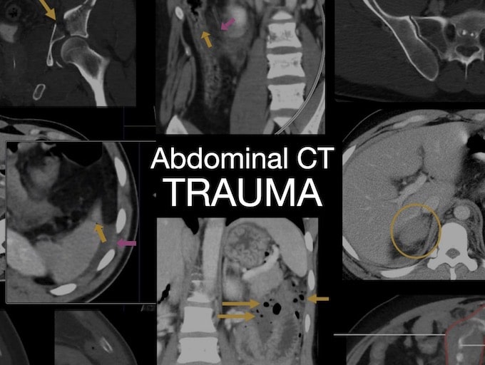
Abdominal CT: Trauma series. Evaluating bowel and mesenteric trauma with examples cases

![]()
Abdominal CT: Trauma series. Evaluating bowel and mesenteric trauma with examples cases
Abdominal CT: Trauma series. Case study to review of the major patterns of solid organ injury identified on abdominal CT.
Abdominal CT: Trauma series. Imaging renal, ureter, and adrenal injuries
Abdominal CT: Trauma series. Diagnosing spleen, liver, and pancreatic injuries
Abdominal CT: Trauma series. Solid organ injuries. Trauma is a leading cause of death worldwide and it has two broad classifications: Blunt and penetrating
Abdominal CT: Identifying intestinal ischaemia. Mesenteric ischaemia can be visualised on CT through examining blood vessels and the bowel
Abdominal CT: Identifying intestinal ischaemia. Mesenteric ischaemia can be visualised on CT through examining blood vessels and the bowel
Abdominal CT: cholecystitis. Cholecystitis is inflammation of the gallbladder commonly caused by an obstruction of the cystic duct by a gallstone
Abdominal CT: Spotting renal infections. Pyelonephritis infection of the urinary tract starts in the bladder involving the kidneys through the ureters
Abdominal CT: renal stones. Renal stones are hard deposits that form in the kidney. They can move down the urinary tract, causing obstruction and pain as well as blood in the urine.
Abdominal CT: pancreatitis. Recognizing acute necrotizing pancreatitis; and late stage development of pseudocysts and walled-off necrosis
Abdominal CT: bowel perforation. Perforation of the gastrointestinal tract can be due to a variety of causes.