![]()
Left Posterior Fascicular Block (LPFB)
Left posterior fascicular block LPFB (left posterior hemiblock), impulses are conducted to the left ventricle via the left anterior fascicle

![]()
Left posterior fascicular block LPFB (left posterior hemiblock), impulses are conducted to the left ventricle via the left anterior fascicle

The ATACC group take us through the evolution of human patient simulation. How can clinicians experience emotional attachment that comes when practicing procedures on rubber human simulators?
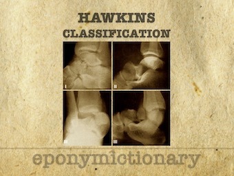
Hawkins classification: Classification system for talar neck fractures. Hawkins originally described Types I-III in 1970 with Canale and Kelly adding Type IV in 1978
A review of the ECG features of right ventricular outflow tract tachycardia (RVOT), a type of VT, with example ECGs.
Isolated low serum Mg levels are associated with atrial depolarisation and ventricular repolarisation abnormalities, predisposing to ventricular arrhythmias
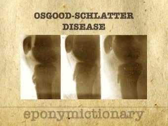
Osgood–Schlatter disease (OSD) Osteochondrosis or traction apophysitis of the tibial tubercle. Paget (1891), Osgood (1903), Schlatter (1903)
What's next after RESCUEicp? -The results of this study may have been disappointing, but there are some questions about the trial itself which we review.
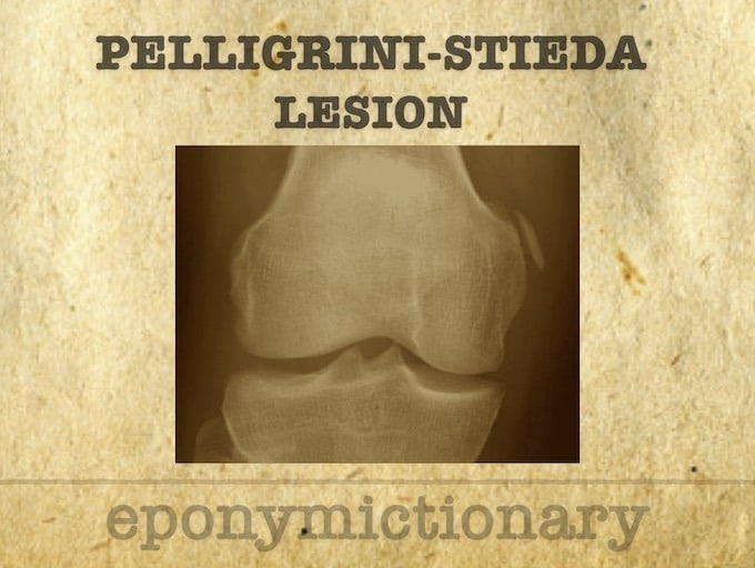
Köhler-Pellegrini-Stieda lesion: ossification near medial femoral collateral ligament adjacent to the margin of the medial femoral condyle.

The story of the Northern Ireland HEMS Service, from the political and public campaign to get it started right through to the teamwork and hard work involved in making this project a reality.
A strong interdependent system of care can improve survival from out of hospital cardiac arrest, with Tony Walker ASM

François Chopart (1743 – 1795) was a French Surgeon. Eponymously associated with Chopart fracture-dislocation, Chopart joint and Chopart amputation.

Critical Care Compendium - The C-IMPLE approach to regional nerve block