![]()
Monteggia fracture
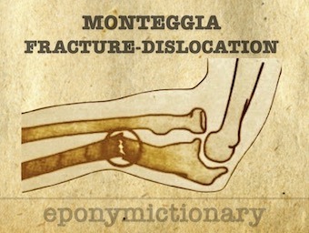
Monteggia fracture. Fracture of the proximal or middle third of the ulna with associated radial head dislocation/instability. GB Monteggia 1812

![]()
Monteggia fracture. Fracture of the proximal or middle third of the ulna with associated radial head dislocation/instability. GB Monteggia 1812

Galeazzi fracture (1934). Fracture of the distal third of the radius with associated Distal radio-ulna joint (DRUJ) disruption.
Naxos disease: a recessively inherited condition with arrhythmogenic right ventricular dysplasia/ cardiomyopathy (ARVD/C) non-epidermolytic palmoplantar keratoderma, and woolly hair
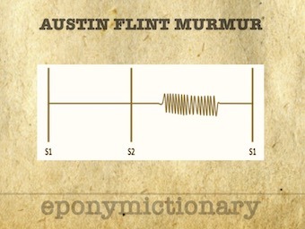
Austin Flint Murmur: Mid diastolic, low pitch rumble heard best at the apex. Absence of opening snap/loud S1 distinguishes from that of mitral stenosis
Primary thrombosis of the subclavian vein at the costoclavicular junction. The formation of an axillo-subclavian vein thrombosis results from endothelial trauma, often as a result of repetitive activity of the upper limbs.

Seldinger Technique a technique for safe percutaneous access to vessels and hollow organs that is widely used today. Sven Ivar Seldinger (1921 – 1998)
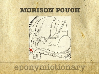
In 1894, Morison published his anatomic description of the hepatorenal space; its role in the surgical treatment of gallbladder disease; and proposed the value of postoperative drainage of that space.
Löffler (Loeffler) syndrome is a transient, self-limiting, and benign pulmonary eosinophilia, characterised by pulmonary opacities on X-ray, elevated blood eosinophils and an acute onset of potential symptoms of mainly cough and dyspnoea.

In 1961, Jack Handyside Barnes, his nine year-old son, and a local surf lifesaver were rushed to Cairns Base Hospital after developing Irukandji syndrome.
Behçet disease: chronic, multisystemic inflammatory condition involving small and large vessels of unknown aetiology. Characterised by the triad of recurrent oral aphthous ulcers, genital ulcers, and iridocyclitis with or without hypopyon.

Charles AHA Bertrand (1777-1849) was a French physician; Least recognised for his self-experimentation with charcoal as an antidote for ingested poisonings.
Rare, acquired clinical syndrome presenting with amenorrhoea, menstrual disorders and reproductive dysfunction secondary to intrauterine adhesions.