![]()

George Chance
George Quentin Chance was an British radiologist. Eponymously associated with the Chance fracture (1948) transverse fracture through a vertebral body

![]()
George Quentin Chance was an British radiologist. Eponymously associated with the Chance fracture (1948) transverse fracture through a vertebral body
Australian virologist Yvonne Cossart (1934–2014), pioneer of parvovirus B19 research, teacher, and reformer of medical education.
James Ramsay Hunt (1874-1937) American neurologist. Renowned for his contributions to the field of neurology. Several conditions bear his name including Ramsay Hunt syndrome (1907)
Howard Henry Tooth (1856–1925), English neurologist; Charcot–Marie–Tooth disease; specialist in spinal degeneration and wartime medical service.
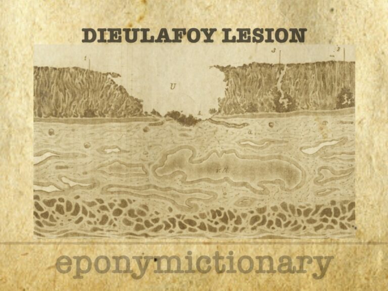
Dieulafoy’s lesion: minute gastric erosion over a large arteriole, causing massive GI bleeding. First defined as exulceratio simplex in 1898.
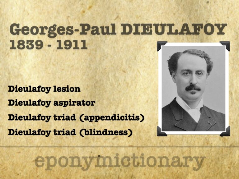
Georges Dieulafoy (1839–1911), French physician, pioneer of gastroenterology; remembered for Dieulafoy lesion, triad(s), and aspirator
Eugen von Bamberger (1858–1921), Austrian internist; co-described Marie–Bamberger syndrome (hypertrophic pulmonary osteoarthropathy); pioneer of clinical diagnostics.
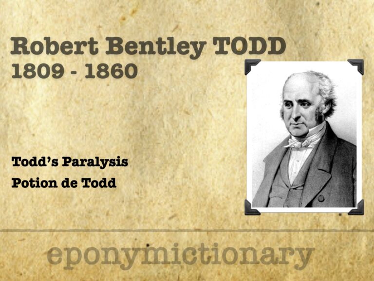
Robert Bentley Todd (1809-1860) was an Irish physician. Provided early depictions of migraine, peripheral neuritis, and postepileptic paralysis (Todd's palsy). He also gave an important discourse on locomotor ataxy (tabes dorsalis).
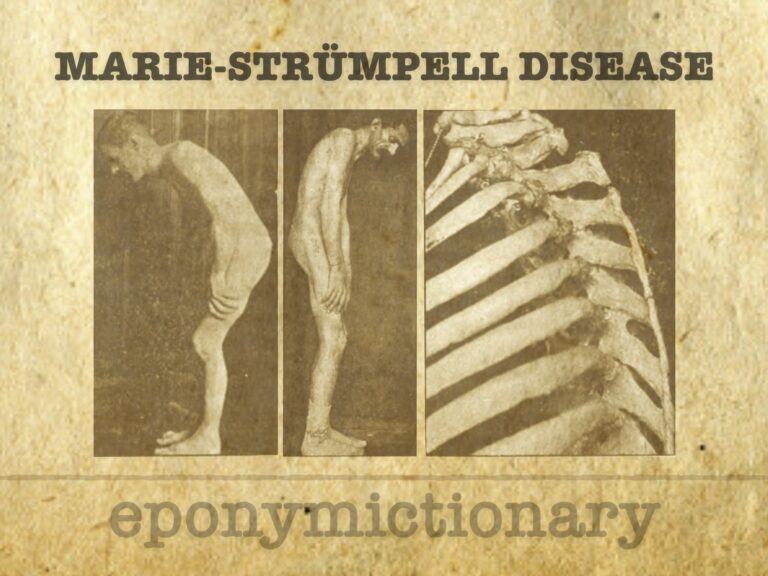
Marie-Strümpell disease (ankylosing spondylitis): a chronic inflammatory spinal arthritis with progressive axial fusion, first described by Pierre Marie and Adolf Strümpell in the late 19th century.
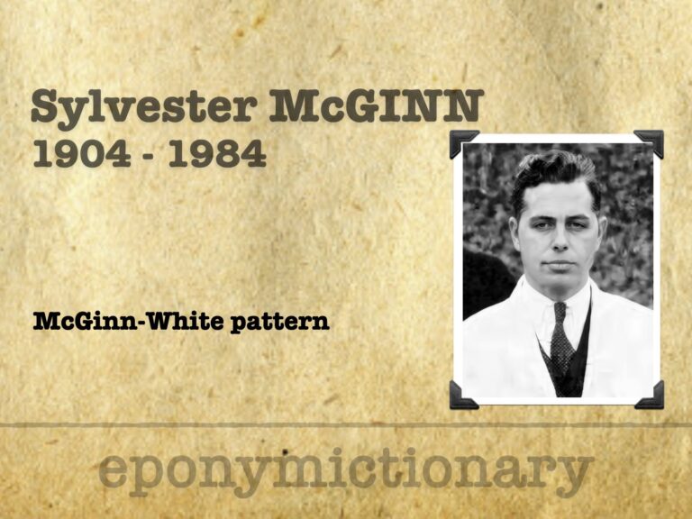
Sylvester McGinn (1904–1984); American cardiologist; McGinn-White sign (S1Q3T3) in pulmonary embolism; pioneer in Boston cardiac care and research.
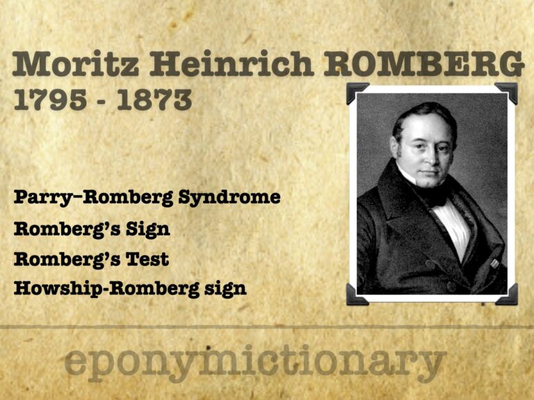
Moritz Heinrich Romberg (1795–1873) a founder of clinical neurology, Romberg’s sign, Parry–Romberg syndrome and Howship–Romberg sign
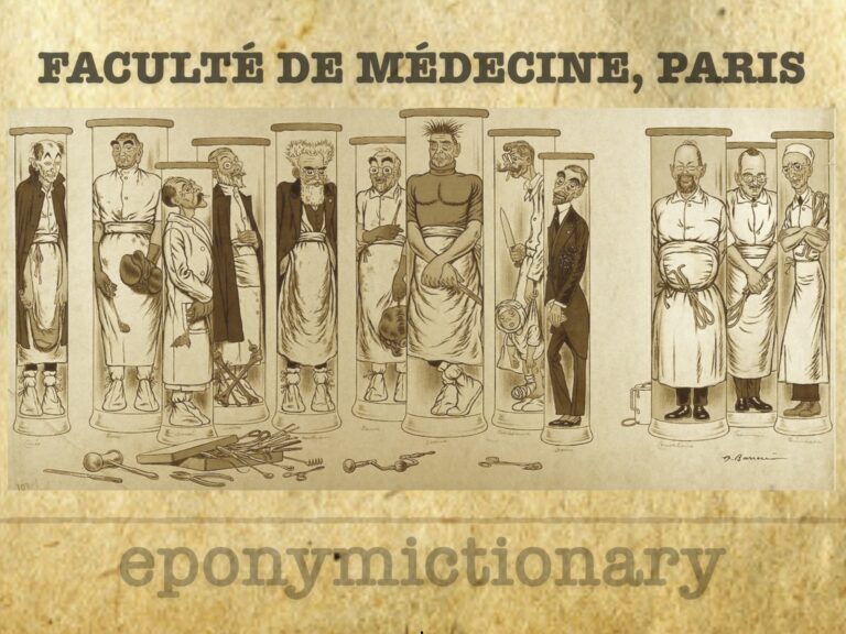
Adrien Barrère (1874-1931) produced a series of lithographs of the Professors in the Faculties of Medicine. Fourth lithograph 'A cluster of surgeons' in 1910